Methylene Blue as Objective Neuroprotective Support in mTBI and cTBI
NeuroLyteDxARC Medical CenterInjury Accident NetworkTop Rated Medical
Mild and concussion-type TBI trigger a secondary injury cascade — mitochondrial dysfunction, oxidative stress, blood–brain barrier disruption, and neuroinflammation — that drives persistent symptoms despite normal imaging. Methylene blue (MB), a USP-grade phenothiazine, acts within the mitochondrion as a redox catalyst to sustain ATP and reduce oxidative stress, and in preclinical models reduces lesion volume, edema, and behavioral deficits. Human evidence remains limited to healthy-volunteer cognition and neurodegenerative disease, with no completed randomized trial in TBI, and MB carries serious risks including serotonin syndrome and contraindication in G6PD deficiency and pregnancy. The paper presents the mechanism, the state of the evidence, a structured intravenous administration framework, and the safety and medico-legal considerations for responsible, investigational, off-label use.
Methylene blue is a mechanistically compelling investigational candidate for limiting the secondary injury cascade following mTBI/cTBI, particularly in personal injury (PI) contexts such as motor vehicle collisions and falls. After trauma, neurons enter a bioenergetic crisis: mitochondria fail, ATP collapses, oxidative stress and neuroinflammation propagate, and the BBB becomes permeable. MB targets several of these processes simultaneously by acting within mitochondrial electron transport pathways.
Despite a coherent mechanism and consistent animal data, MB for TBI remains investigational and off-label. The strongest controlled human evidence comes from a randomized, placebo-controlled functional-MRI study in healthy adults, not injured patients, in which a single low dose improved memory retrieval and increased task-related brain activity (this paper addresses intravenous administration; see Section 6). The clinical and medico-legal value of MB therefore depends on accurate characterization: claims must reflect the evidence, and use must respect strict safety boundaries.
This paper proposes a practical framework for responsible investigational use while emphasizing that efficacy in human mTBI has not yet been established.
In injury, accident, and expert-witness settings, MB should be presented as adjunctive and investigational, never as established concussion monotherapy. The most important practical safeguard is medication reconciliation: depression and anxiety are common after concussion and are frequently treated with SSRIs/SNRIs, which makes the serotonergic interaction a foreseeable and potentially fatal hazard in this exact population.
Properly framed, MB can be discussed in treatment plans and future-cost projections as an emerging, mechanism-based option, provided the documentation states its off-label status, screens for contraindications, records informed consent, and attaches the safety caveats. Overstating the evidence undermines credibility; understating the mechanism overlooks a plausible avenue of care. Accurate characterization protects both the patient and the defensibility of the report.
Traumatic brain injury is among the most common neurological injuries in clinical practice. In the United States alone, an estimated several million people sustain a TBI each year, the great majority classified as mild, yet a substantial minority go on to experience symptoms, cognitive slowing, headache, dizziness, and mood change, that persist for months or longer. Despite this burden, there is at present no FDA-approved pharmacologic agent that halts or reverses the underlying secondary injury cascade; standard care remains largely supportive and symptom-directed. This therapeutic gap is the backdrop against which mitochondria-targeted agents such as methylene blue have drawn interest.
Methylene blue (methylthioninium chloride) is a tricyclic phenothiazine dye first synthesized in 1876, long used clinically to treat methemoglobinemia and as a surgical stain. Chemically it is a redox (reduction–oxidation) catalyst: at low concentrations it readily accepts and donates electrons, which underlies its activity inside mitochondria. MB crosses the BBB efficiently, after intravenous administration its brain concentration can substantially exceed plasma levels within roughly an hour, making it well suited to act on injured neural tissue.
Traumatic brain injury unfolds in two phases. Primary injury is the immediate mechanical damage from the impact or jolt. Secondary injury develops over hours to months and is where most therapeutic opportunity lies: mitochondrial dysfunction, ATP failure, oxidative stress, excitotoxicity, BBB breakdown, cerebral edema, cellular shearing, and microglia-driven neuroinflammation. Mild TBI and concussion sit at the lower-severity end of this spectrum yet can still produce persistent cognitive slowing, attention and memory difficulty, headache, and mood change.
A central frustration in concussion care is the scarcity of proactive pharmacologic treatments that limit the secondary cascade. This therapeutic gap has driven interest in mitochondria-targeted agents such as MB. However, enthusiasm in wellness and nootropic communities has outpaced controlled human evidence, and dosing guidance frequently originates from commercial sources rather than peer-reviewed TBI trials. This paper distinguishes mechanism from outcome, separates preclinical from human data, and provides a structured framework for any investigational use.
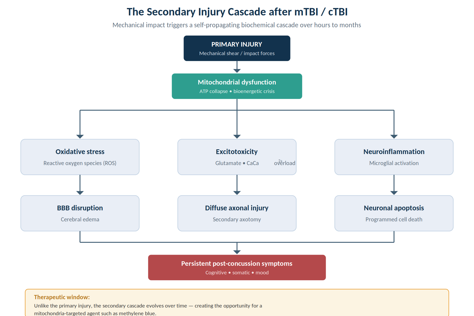
MB's candidacy in TBI rests on overlapping mechanisms, each countering a distinct part of the secondary injury cascade. The table below maps mechanism to injury process.
| Mechanism of methylene blue | Secondary-injury process it counters |
|---|---|
| Mitochondrial electron shuttling → sustained ATP | Energy (ATP) failure and bioenergetic collapse in injured neurons |
| Antioxidant activity at low dose; NRF2 support | Oxidative stress and free-radical damage |
| Promotion of autophagy | Accumulation of damaged proteins/organelles; degeneration |
| Inhibition of microglial activation | Neuroinflammation |
| Reduced edema; preserved BBB; lower S100 | Cerebral edema and BBB breakdown |
| Reduced neuronal apoptosis | Programmed cell death following injury |
Mitochondrial dysfunction is a hallmark of brain injury, and mitochondria are a specific therapeutic target after TBI. By rerouting electrons in the electron transport chain, MB can boost ATP production, enhance oxygen consumption, and improve cellular energetics, helping injured-but-viable neurons survive the energy crisis. At low concentrations MB behaves as an antioxidant and has been linked to activation of the NRF2 antioxidant-defense pathway. Critically, this effect is biphasic and dose-dependent: above approximately 4 mg/kg MB can flip to a pro-oxidant and cause harm. The therapeutic window is narrow, and "more" is not better.
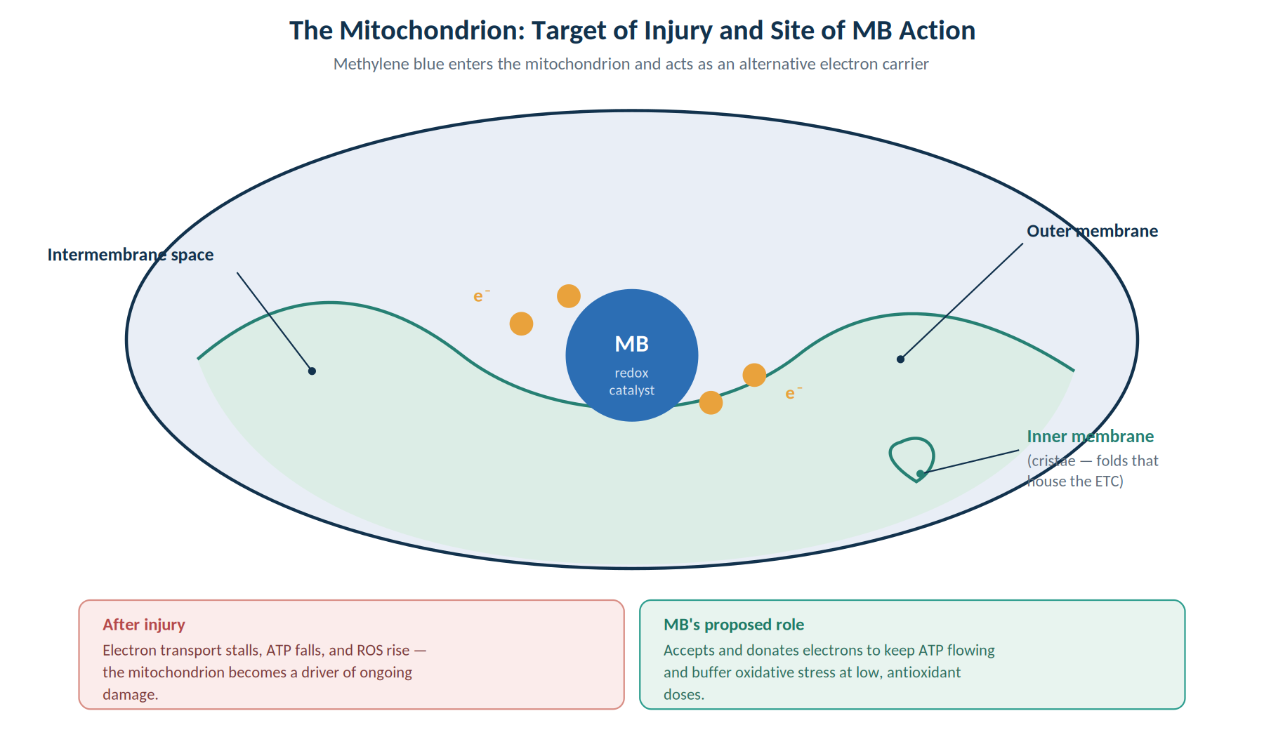
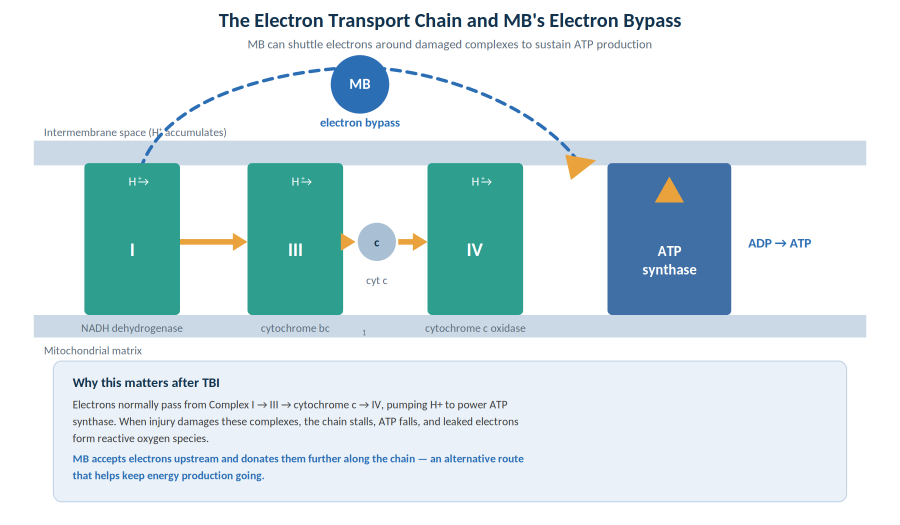
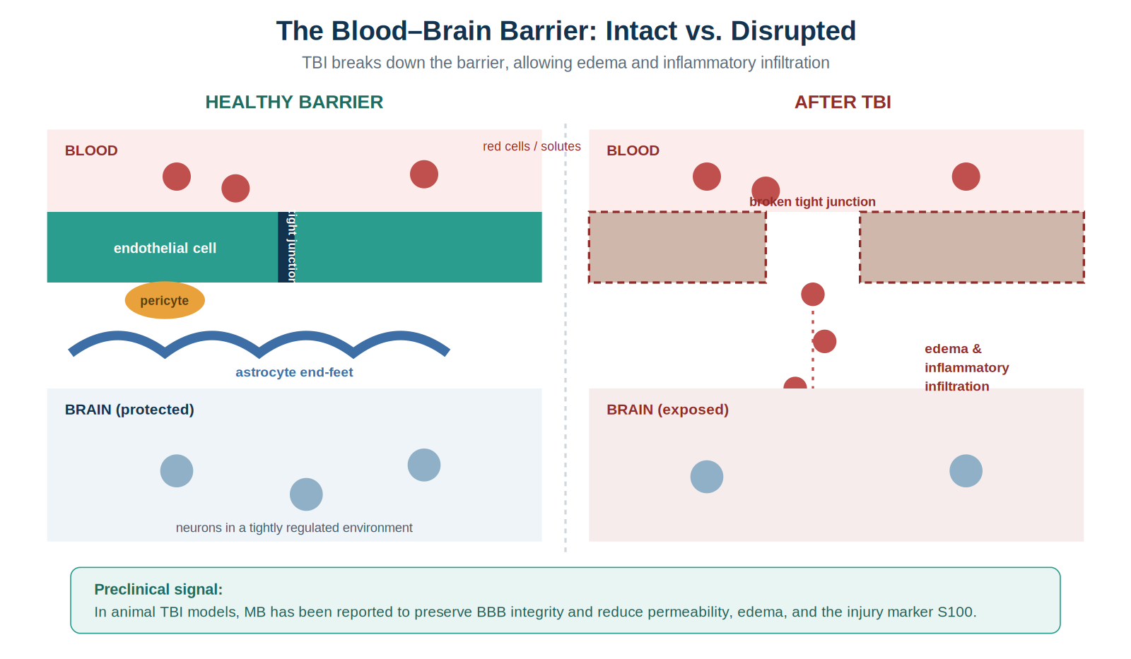
In a controlled cortical impact mouse model, MB exerted neuroprotection by promoting autophagy and inhibiting microglial activation, with benefit in both acute (24–72 h) and chronic (14-day) phases. Separate rat work showed that a single intravenous dose given shortly after injury reduced neurological deficit, preserved BBB integrity, and lowered expression of the injury marker S100, while protecting cultured neurons from inflammatory (lipopolysaccharide) toxicity. Other studies report reduced cerebral edema, attenuated microglial activation, reduced neuronal apoptosis, and improved behavioral recovery in animal models, including acute depressive-like behavior.
A pathology central to concussion that the preceding mechanisms only partly capture is the uncoupling of cerebral blood flow (CBF) from metabolic demand. After concussion, the brain commonly enters a state of cerebral hypoperfusion and neurovascular uncoupling: the normally tight matching of regional blood flow to local energy needs is disrupted, so that oxygen and glucose delivery no longer track neuronal activity. Because mitochondrial dysfunction simultaneously impairs the brain's ability to extract and use the oxygen that does arrive, the injured brain faces a combined supply-and-utilization deficit, a plausible substrate for persistent post-concussion symptoms.
MB is mechanistically relevant to this axis because it acts on both sides of the flow–metabolism relationship. Through its action at mitochondrial cytochrome c oxidase and Complexes I–III, MB can enhance cerebral oxygen utilization and the cerebral metabolic rate of oxygen (CMRO₂) and glucose, improving the efficiency with which delivered substrate is converted to ATP. Preclinical neuroimaging has shown that low-dose MB can increase regional glucose uptake, CBF, and CMRO₂ and can potentiate stimulus-evoked responses, consistent with strengthened flow–metabolism coupling.
An important caveat: the human and dose-response data are mixed. While rodent work has shown parallel increases in CBF and oxygen metabolism, a more recent human and rat neuroimaging study reported that acute MB instead reduced global CBF and CMRO₂, an effect attributed in part to MB's inhibition of nitric-oxide–mediated vasodilation, which becomes more pronounced at higher doses. These findings underscore the hormetic, dose-dependent nature of MB's cerebrovascular effects and caution against assuming uniform blood-flow benefit. The therapeutic hypothesis, that low-dose MB improves metabolic efficiency and oxygen utilization in a hypometabolic, uncoupled post-concussion brain, remains plausible but unproven in injured humans.
Diffuse axonal injury (DAI) is arguably the central pathology of concussion: rotational and acceleration–deceleration forces stretch and shear white-matter axons, producing widespread disconnection that conventional static imaging frequently misses. Crucially, axons are usually not severed at the moment of impact. Instead, injury triggers a progressive, energy-dependent cascade, secondary axotomy, that unfolds over hours to days and is, in principle, a therapeutic target.
The axonal cascade is fundamentally a bioenergetic failure:
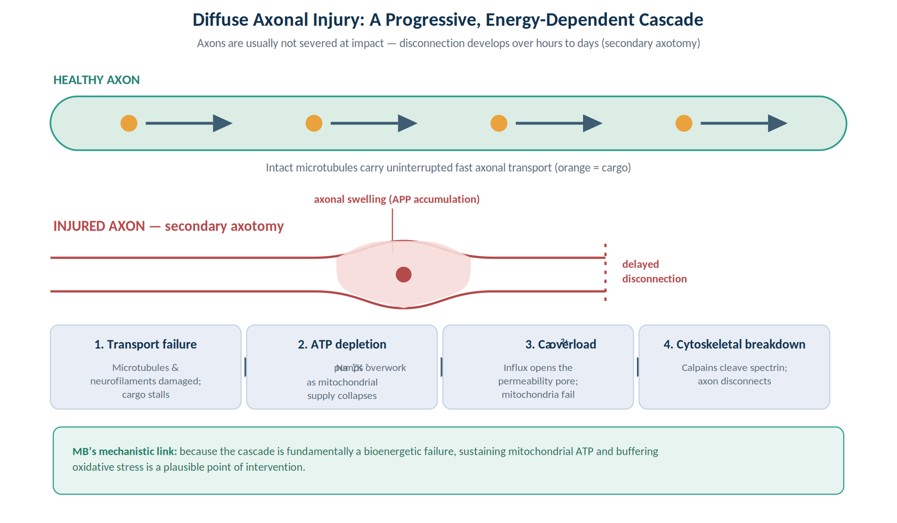
MB intersects this cascade at its bioenergetic core. By sustaining mitochondrial electron transport and ATP production, buffering the oxidative stress that accompanies calcium overload, and supporting mitochondrial integrity, MB could in theory help injured-but-viable axons meet the energy demands of secondary axotomy and avoid progression to disconnection. This is a mechanistic rationale, not a demonstrated effect: no study has shown that MB preserves axonal integrity or improves white-matter outcomes in human DAI. It does, however, connect MB directly to the pathology most responsible for concussion's persistent cognitive and connectivity deficits.
It is essential to separate preclinical from human evidence. The animal literature is substantial and consistent; the human TBI literature is essentially absent at the level of controlled outcome trials. The strongest human cognitive data come from healthy volunteers, not injured patients.
The pivotal controlled human study is a randomized, double-blind, placebo-controlled functional-MRI trial in 26 healthy adults (ages 22–62). A single low dose of USP-grade MB (280 mg, approximately 4 mg/kg, administered orally in that study) increased fMRI activity during sustained-attention and short-term-memory tasks and was associated with roughly a 7% increase in correct responses during memory retrieval. MB at 4 mg/kg has been used safely for up to a year in clinical-trial settings, and a phase II program in Alzheimer's disease (the methylthioninium agent "Rember") reported slowed progression versus placebo. These cognitive trials used oral dosing; the cerebral metabolic and hemodynamic studies in Section 3.3 used intravenous MB, which is the route addressed by this paper.
The crucial limitation: these human findings are in healthy volunteers and in neurodegenerative disease, not in mTBI/cTBI patients. No completed randomized controlled trial demonstrates that MB improves recovery, symptom burden, or function after human traumatic brain injury. The TBI rationale in humans remains an extrapolation from mechanism plus animal data plus healthy-subject cognition.
| Domain | Strength | What it shows |
|---|---|---|
| Mechanism | Strong / coherent | Mitochondrial, antioxidant, anti-inflammatory actions align with TBI pathophysiology |
| Animal TBI | Moderate–strong | Consistent reductions in lesion, edema, BBB damage, inflammation, deficits |
| Human cognition (healthy) | Moderate | One RCT: improved memory retrieval and task-related brain activity |
| Human TBI / concussion | Insufficient | No completed outcome RCT; rationale is extrapolated |
Safety is the decisive practical issue with MB, arguably more than efficacy. Its most dangerous property is potent MAO inhibition.
MB is a potent MAO-A inhibitor and is structurally related to tricyclic antidepressants. Combined with serotonergic drugs, it can cause serotonin syndrome, which has been fatal in reported cases. The FDA has issued a drug-safety communication on this interaction. Implicated agents include SSRIs, SNRIs, MAOIs, tricyclic antidepressants, triptans, tramadol, dextromethorphan, lithium, and other serotonergic drugs. Because depression and anxiety are common after concussion and frequently treated with SSRIs/SNRIs, this interaction is especially relevant in a post-injury population and demands careful medication reconciliation.
Only pharmaceutical (USP) grade methylene blue should be used in humans. Industrial or laboratory-grade material may contain heavy-metal contaminants (e.g., arsenic, cadmium, mercury, lead). A Certificate of Analysis documenting high purity should be obtained for any product intended for human use.
This section addresses intravenous (IV) administration only. Oral, sublingual, and other self-administered routes are outside the scope of this paper and are not recommended for the TBI/concussion context: IV delivery occurs under direct medical supervision, allows precise dose control and monitoring, and avoids the variable absorption and unsupervised use associated with consumer oral products. A critical caveat governs everything below: there is no validated human IV dosing regimen for methylene blue in TBI or concussion. The parameters that follow are extrapolated from the established IV pharmacology of MB (its FDA-approved use in methemoglobinemia), from preclinical TBI models that used IV dosing, and from clinical-pharmacology references; they are informational, not prescriptive.
Potential candidates are adults with physician-diagnosed mTBI/cTBI being considered for adjunctive, off-label IV neuroprotective support under direct medical supervision. Potential candidates may include:
G6PD screening before IV MB is the single most important pre-treatment safeguard.
IV methylene blue should be administered only in a closely monitored clinical setting equipped to manage hypersensitivity reactions and serotonergic toxicity, with vital signs, ECG, and (in the methemoglobinemia context) methemoglobin levels monitored during and after administration. Because of the MAOI interaction, IV MB should never be given without prior medication reconciliation and, when time permits, G6PD screening.
MB follows a hormetic (biphasic) dose–response: low doses are antioxidant and beneficial, while higher doses become pro-oxidant and can paradoxically worsen methemoglobinemia, with pro-oxidant harm reported above approximately 4 mg/kg and methemoglobinemia risk above approximately 7 mg/kg. The therapeutic window is narrow, and "more" is not better. As an illustration, one investigational clinical approach administers methylene blue using a low fixed-dose ladder of 25 mg, 50 mg, 75 mg, and 100 mg per dose, titrated to symptom severity and tolerability, and not to exceed 150 mg per dose. Depending on symptom severity, methylene blue may be dosed twice weekly for more severe presentations or once weekly for milder presentations. These low fixed doses sit well below the toxicity thresholds above, and the lowest effective dose should always be used. The table below summarizes the fixed-dose ladder.
| Dose (fixed) | Rate / notes | Context |
|---|---|---|
| 25 mg | Slow IV over 5–30 min | Lowest starting dose; mild presentations or initial titration |
| 50 mg | Slow IV over 5–30 min | Step-up dose as tolerated and indicated by symptom severity |
| 75 mg | Slow IV over 5–30 min | Step-up dose for more severe or refractory presentations |
| 100 mg | Slow IV over 5–30 min | Upper routine dose; do not exceed 150 mg in a single dose |
Important — origin of these dose figures. These dose ranges represent one clinic's conservative investigational protocol, developed from established intravenous methylene blue pharmacology, the toxicology literature, and published preclinical neuroprotection studies. They are not published, validated, evidence-based dosing recommendations for TBI, and should not be interpreted as such. The fixed dose is selected by the prescriber according to symptom severity and tolerability and is not to exceed 150 mg per dose.
Practical principle. Use the lowest effective dose under monitoring, give it slowly, do not exceed the 150 mg per-dose maximum, and frame any TBI application as adjunctive and investigational, individualized by a prescriber, and contingent on the exclusions in 6.1.
The prescriber selects a fixed dose of 25, 50, 75, or 100 mg (not to exceed 150 mg) according to symptom severity and tolerability. To prepare, divide the selected milligram dose by the stock concentration shown on the product or compounding label to obtain the volume to withdraw:
Stock volume (mL) = dose (mg) ÷ stock concentration (mg/mL)
The withdrawn stock is then added to 50 mL of D5W and infused over 5–30 minutes. Worked example: a 50 mg dose drawn from a 10 mg/mL reconstituted stock is 50 mg ÷ 10 mg/mL = 5 mL, diluted in 50 mL D5W (final volume ≈ 55 mL), infused slowly. The table gives withdrawal volumes for each fixed dose at the two most common stock concentrations; always confirm the actual concentration on the label before calculating.
| Fixed dose | Volume from 5 mg/mL stock | Volume from 10 mg/mL stock | Diluent for infusion |
|---|---|---|---|
| 25 mg | 5 mL | 2.5 mL | 50 mL D5W |
| 50 mg | 10 mL | 5 mL | 50 mL D5W |
| 75 mg | 15 mL | 7.5 mL | 50 mL D5W |
| 100 mg | 20 mL | 10 mL | 50 mL D5W |
Diluent should be D5W (or sterile water), not 0.9% sodium chloride. Do not exceed 150 mg in a single dose. These calculations are provided for preparation accuracy; they reflect one clinic's fixed-dose schedule, are not a validated TBI treatment protocol, and must be confirmed against current product labeling and institutional policy.
Section 6 limitation. The dosing, dilution, and preparation information presented in this section is derived from FDA-approved methylene blue pharmacology, clinical-pharmacology references, and preclinical neuroprotection studies. It should not be interpreted as an established treatment protocol for mTBI/cTBI. Clinical decisions remain the responsibility of the treating provider.
The following algorithm summarizes the investigational decision pathway described in this section, from presentation through screening, contraindication check, informed consent, IV administration, and objective outcome tracking.
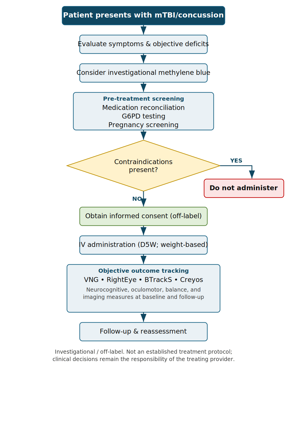
In an injury, accident, or expert-witness context, claims about MB should be framed precisely to reflect the evidence and avoid overstatement.
Because MB is investigational and carries a narrow safety margin, candidacy is deliberately restrictive rather than universal, and the reasons a given patient does or does not receive it should be documented. Patients are considered only when they have a physician-diagnosed mTBI/cTBI with objective, measurable deficits and persistent symptoms despite conservative care, and when they clear the contraindication screen in Section 6.1. Conversely, patients are excluded when any absolute contraindication is present, most commonly current serotonergic medication, which is frequent in a post-concussion population treated for mood or anxiety, or when G6PD status or pregnancy cannot be confirmed. Making this selection logic explicit in the record protects the patient, explains apparent variation in who is and is not offered MB, and strengthens the defensibility of any report.
Given the investigational nature of methylene blue for mTBI/cTBI, informed consent should specifically document:
Methylene blue is one of the more mechanistically compelling investigational candidates for limiting the secondary injury cascade after mTBI/cTBI. It acts where the injury does the most ongoing damage, within mitochondrial electron transport pathways, and across animal models it reliably reduces edema, BBB disruption, neuroinflammation, apoptosis, and behavioral deficits. A controlled human study shows it can enhance memory and attention-related brain activity in healthy adults at low doses, and it has an unusually long pharmaceutical track record.
What is missing is the part that matters most clinically: controlled trials in actual mTBI/cTBI patients with meaningful functional and symptom endpoints. Until those exist, MB in TBI should be presented as a promising, investigational, off-label option, never as established therapy, and always within strict safety boundaries: serotonergic-drug screening, G6PD and pregnancy exclusion, conservative dosing, and pharmaceutical-grade product.
Although most MB–TBI research addresses the acute phase, the population most likely to seek out this paper consists of patients with persistent symptoms, at 3 months, 6 months, 1 year, or longer after injury. Chronic post-concussion syndrome (PCS) is increasingly understood not as a purely psychological sequela but as a state with measurable biological correlates that overlap substantially with MB's mechanisms:
No controlled human evidence currently exists for MB in chronic PCS. The rationale is entirely mechanistic: the biology of persistent PCS (chronic neuroinflammation, mitochondrial dysfunction, hypometabolism, and connectivity loss) maps onto MB's known actions, and preclinical work suggesting that durable benefit may require repeated dosing is at least consistent with a chronic-phase treatment model. This alignment makes chronic PCS a logical target for future trials but does not justify clinical claims of benefit today.
Importantly, these correlates are not transient. Chronic neuroinflammation, chronic cerebral hypometabolism, and persistent white-matter dysfunction have each been documented years after the initial injury, connecting chronic PCS directly to the diffuse axonal injury described in Section 3.4: the white-matter disconnection that begins as secondary axotomy can leave durable structural and metabolic signatures long after symptoms might otherwise be dismissed as functional. A treatment hypothesis aimed at the chronic phase must therefore target this persisting biology rather than the acute cascade alone.
If MB is ever studied or used investigationally in this population, response should be tracked with objective, reproducible measures rather than symptom report alone, consistent with a comprehensive functional neurological evaluation (C-FNE) framework. Candidate measures that could quantify change over a treatment course include:
Pairing any investigational MB use with this kind of objective monitoring would allow response to be demonstrated rather than assumed, strengthen both clinical decision-making and medico-legal defensibility, and generate the structured data the field currently lacks.
A concise summary of the paper's operational message:
References are numbered in citation-sequence order (order of first appearance in the text) so that each in-text superscript points to the source it supports.
Adenosine Triphosphate (ATP): The primary energy-carrying molecule used by cells. ATP depletion is a hallmark of the secondary injury cascade following TBI.
Blood–Brain Barrier (BBB): A specialized network of endothelial cells that regulates movement of substances between the bloodstream and the CNS. Disruption after TBI may contribute to edema, neuroinflammation, and neuronal dysfunction.
Concussion: A form of mild TBI caused by a bump, jolt, or blow to the head, or forces transmitted to the head, resulting in temporary disruption of normal brain function.
Diffuse Axonal Injury (DAI): Microscopic white-matter injury from rotational or acceleration–deceleration forces that stretch or shear axons; a primary pathological mechanism underlying concussion and TBI.
Electron Transport Chain (ETC): A series of inner-mitochondrial-membrane protein complexes that generate ATP through electron transfer and oxidative phosphorylation.
Glucose-6-Phosphate Dehydrogenase (G6PD) Deficiency: An inherited enzymatic disorder increasing susceptibility to hemolytic anemia. MB is contraindicated in G6PD deficiency.
Hormesis (Hormetic Dose Response): A biphasic response in which low doses produce beneficial effects while higher doses may produce toxicity or harm.
Methemoglobinemia: A condition in which hemoglobin is oxidized to methemoglobin, reducing oxygen transport. MB is FDA-approved for acquired methemoglobinemia.
Methylene Blue (MB): A synthetic phenothiazine that functions as a redox catalyst and mitochondrial electron carrier. FDA-approved for methemoglobinemia; investigated for neuroprotection.
Mild Traumatic Brain Injury (mTBI): A brain injury from external mechanical forces producing transient neurological dysfunction (e.g., headache, dizziness, cognitive impairment, visual disturbance, balance dysfunction, mood change).
Mitochondrial Dysfunction: Impaired mitochondrial function causing reduced ATP production, increased oxidative stress, and decreased cellular energy availability.
Monoamine Oxidase Inhibitor (MAOI): A compound that inhibits monoamine oxidase enzymes metabolizing serotonin, dopamine, and norepinephrine. MB possesses MAOI activity and may interact dangerously with serotonergic drugs.
Neuroinflammation: The CNS inflammatory response involving microglia, astrocytes, and inflammatory signaling. Persistent neuroinflammation is implicated in chronic post-concussion symptoms.
Neuroprotection: Interventions intended to preserve neuronal structure and function by reducing secondary injury mechanisms such as oxidative stress, inflammation, apoptosis, and mitochondrial dysfunction.
Objective Neurological Assessment: The use of measurable physiological, cognitive, visual, vestibular, balance, or neuropsychological testing to evaluate function independent of self-report.
Off-Label Use: Use of an FDA-approved medication for an indication, dose, route, or population not specifically approved by the FDA.
Oxidative Stress: An imbalance between reactive oxygen species production and antioxidant defenses, resulting in cellular damage.
Randomized Controlled Trial (RCT): A study design in which participants are randomly assigned to treatment or control groups to evaluate efficacy and safety.
Reactive Oxygen Species (ROS): Highly reactive oxygen-containing molecules that can damage proteins, lipids, DNA, and cellular structures when present in excess.
Redox (Reduction–Oxidation) Reaction: A reaction involving electron transfer between molecules. Mitochondrial energy production depends on redox reactions, and MB acts as a redox catalyst.
Secondary Injury Cascade: The biochemical and cellular events following the initial injury, mitochondrial dysfunction, ATP depletion, oxidative stress, neuroinflammation, edema, BBB disruption, and apoptosis, which may remain active for days to months and is a major therapeutic target.
Serotonin Syndrome: A potentially life-threatening condition caused by excessive serotonergic activity, with agitation, hyperthermia, tremor, autonomic instability, and altered mental status.
This document is an educational and scientific review intended for clinicians and informed readers. It is not medical advice, does not establish a standard of care, and is not a treatment protocol. Methylene blue is not FDA-approved for the treatment of traumatic brain injury; all neuroprotective and cognitive uses described here are investigational or off-label. Decisions for any individual patient must be made by a qualified prescriber after review of contraindications, concurrent medications, and current evidence.