Concussion-type traumatic brain injury commonly produces measurable neurological dysfunction that CT and conventional MRI do not detect, while symptom inventories remain nonspecific and cannot localize dysfunction to a system. The Comprehensive Functional Neurological Evaluation (C-FNE™) sequences instrument-derived measurement, clinician-administered testing, validated patient-reported measures, and documented activity and participation limitation into one auditable examination spanning nine functional domains. The paper sets out the examination architecture, the rules governing incomplete and technically invalid components, the cross-domain interpretation process, and the relationship to the Short-Term Concussion Disability (ST-CD) Scale. It is presented as a structured clinical methodology, not a validated diagnostic test — it does not diagnose concussion, encode causation, determine effort, or establish permanent impairment — and a six-phase validation roadmap remains to be completed.
Terminology statement. Throughout this paper, concussion-type traumatic brain injury (cTBI) is used as a descriptive term for the clinical entity most often labeled concussion or mild traumatic brain injury. It is used because the population addressed here is defined by injury phenotype and functional presentation rather than by an initial severity index, and because the word mild poorly conveys the functional burden many of these patients carry. The term is descriptive only. It is not proposed as a replacement for any existing diagnostic classification, is not a distinct nosologic entity, and does not alter established diagnostic criteria. Section 1.1 deliberately retains concussion and mild traumatic brain injury where the source literature and established equivalence require it.
Executive Summary
This summary is written for clinicians, attorneys, insurers, and health-system administrators who need the framework’s scope and limits without the full methodology. Every statement below is developed and qualified in the body of the paper.
What it is. The Comprehensive Functional Neurological Evaluation (C-FNE™) is a standardized, sequenced clinical examination that measures neurological function across nine domains in a single outpatient encounter and reports every component result in its native units alongside the reference range applied. It is a procedure and a documentation method. It is not a device, not a questionnaire, and not a test with a single score.
The problem it addresses. After cTBI, the clinically dominant abnormalities are disturbances of function — vestibular, oculomotor, postural, cognitive, autonomic, and affective — in patients whose conventional imaging is normal. The objective tools that can measure those disturbances exist, but they are ordered separately, performed in different settings on different days, and reported in incompatible formats. The resulting chart contains a great deal of data and very little integrated interpretation.
What it contains. Eight core components — history and symptom assessment, cranial nerve screening, fourcondition instrumented balance testing, computerized cognitive assessment, psychological and sleep screening, quantitative strength testing, videonystagmography with oculomotor testing, and a structured functional participation interview — supplemented by conditional components performed when specifically indicated. Findings are organized into nine functional domains.
What it produces. A single report containing the raw measured values, the reference ranges applied, the validity status of every component, an explicit designation for anything not tested or not interpretable, a domain-by-domain conclusion, and a cross-domain synthesis. The report is designed so that an independent reviewer can reconstruct each domain conclusion from the underlying data. Where the examination is sufficiently complete, its outputs may be converted into a Short-Term Concussion Disability (ST-CD) Scale profile.
What it supports today. Characterizing which neurological systems are functioning abnormally in an individual patient; directing treatment and referral to the specific domains shown to be impaired; documenting objectively measured findings in a form that can be audited; and providing a structured, repeatable basis for describing present function in clinical, administrative, and legal settings.
What it does not support. The C-FNE does not diagnose concussion — diagnosis is clinical and event-anchored. It does not establish event-specific causation, does not assess effort, malingering, or credibility, does not establish permanent impairment, and does not determine entitlement to benefits or compensation. A normal examination does not exclude a prior injury, and an abnormal examination does not by itself attribute the abnormality to any particular event.
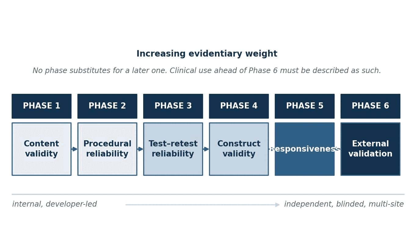
What validation remains. The individual component tests are independently developed and separately published. The integrated framework has not completed formal content-validity, procedural- and inter-rater-reliability, test–retest, construct-validity, responsiveness, or independent external-replication studies. It therefore has no published error rate, no established minimal detectable change, and no independent replication. Until the six-phase validation program in Section 23.0 is complete, the framework should be described as a structured clinical methodology with documented procedures and unestablished measurement properties. That description is accurate and is not a criticism; it is the position occupied by many widely used clinical assessment frameworks. What is not defensible is describing it otherwise.
Intended use. The C-FNE is intended for the standardized characterization of current functional neurological findings in adults undergoing evaluation after suspected or clinically diagnosed cTBI. It is intended to support domain-specific treatment planning, referral, serial clinical documentation, and communication of examination findings between clinicians. It is not intended for emergency triage, for independent diagnosis, for determination of event-specific causation, for assessment of effort or credibility, for permanent impairment rating, or for stand-alone legal decision-making. Use outside these purposes is outside the scope of the methodology described in this paper and is not supported by its developers.
Minimum reporting dataset. Every C-FNE report identifies the examining personnel and the interpreting clinician; the examination date and the interval since injury; the protocol version; the identity and software version of every instrument used; the test conditions and any deviation from the standard sequence; the measured value of every component in its native units; the reference range applied to each; the completion status of every component, including anything not indicated, not tested, aborted, technically invalid, or clinically uninterpretable, each with its reason; the confounders considered; the conclusion reached for each of the nine domains; and the signature of the clinician who takes responsibility for the integrated interpretation. A report that omits any of these is not a complete C-FNE report. This requirement is developed in Sections 18.4 and 18.5 and specified in full in Table 24.
Introduction
Clinical background
Traumatic brain injury (TBI) is among the most common neurological conditions worldwide, and a modelling study of global burden estimated that approximately sixty-nine million individuals sustain a TBI each year, of whom roughly fifty-six million sustain injuries classified as mild.1 Population-based work from the World Health Organization task force found that seventy to ninety percent of all treated brain injuries are mild and placed the incidence of hospitaltreated mild traumatic brain injury (mTBI) at roughly one hundred to three hundred per one hundred thousand population, while concluding that the true population-based rate is probably above six hundred per one hundred thousand because many injured people never present at all.2 In the United States, surveillance data continue to show head injury as a leading contributor to emergency-department utilization across the age spectrum, with motor vehicle collisions, falls, assaults, occupational events, and sport accounting for the bulk of the burden.3
The prevailing clinical expectation — that the great majority of patients recover within days to a few weeks — is broadly accurate but incomplete. Prospective cohort work has repeatedly identified a substantial minority who remain symptomatic and functionally limited months after injury. In the TRACK-TBI cohort, fifty-three percent of patients with mTBI reported injury-related functional limitation twelve months after injury, including forty-nine percent of those whose head computed tomography was normal — a higher proportion than in an orthopedic-injury comparison group.4 The WHO prognosis review reached a similar conclusion from an earlier evidence base, noting that a nontrivial fraction of adults report persistent symptoms and functional restriction well beyond the expected recovery window.5 It is precisely this population — symptomatic, functionally limited, and structurally normal on imaging — that presents the greatest assessment difficulty and that the framework described here was built to serve.
Two features of this presentation drive that difficulty. The first is that the clinically dominant abnormalities are functional rather than structural. The clinical picture after cTBI is dominated by disturbances of vestibular function, gaze control and visual tracking, postural stability, cognitive efficiency, sleep, autonomic regulation, and mood — that is, by disordered performance of neural systems whose gross anatomy is intact.6,7 These are, by definition, abnormalities of process, and processes must be measured by performance rather than inspected by imaging.
The second is that conventional structural imaging is normal in most patients with uncomplicated cTBI. Head computed tomography is performed to exclude a surgical lesion, not to characterize concussion, and it is negative in the great majority of cTBI presentations.8 Conventional magnetic resonance imaging adds sensitivity for small traumatic lesions and improves outcome prediction at the group level, but remains normal in most patients with persistent symptoms.9,10 Diffusion tensor imaging and related advanced sequences have demonstrated group-level white-matter differences after mTBI, but systematic critical review has found the direction, location, and magnitude of those findings inconsistent across studies and insufficiently characterized at the individual-patient level to support clinical diagnostic use.11,12 A normal conventional scan therefore provides limited information regarding the presence, type, or magnitude of functional neurological impairment, and clinicians and patients alike are frequently misled by the implication that it excludes one.
Reliance on symptoms alone is the usual fallback, and it is inadequate for three separate reasons. Symptoms after cTBI are highly nonspecific: the same cluster of headache, fatigue, poor concentration, irritability, sleep disturbance, and dizziness occurs at appreciable base rates in healthy adults13, in patients with depression, anxiety, chronic pain, and other psychiatric diagnoses14, and in orthopedic-injury comparison groups without head trauma.15 Meta-analytic work on neuropsychological outcome after mTBI has similarly found group-level effects that are small and timedependent in unselected samples, and substantially larger in clinic-referred and litigating samples — a divergence that itself argues against treating any single measure as diagnostic.16 Symptoms are also self-limited in the information they carry about mechanism: a patient who reports dizziness has not thereby told the clinician whether the problem lies in the peripheral labyrinth, the central vestibular pathways, the oculomotor system, orthostatic regulation, cervical proprioception, medication effect, or anxiety — and those possibilities have entirely different treatments. Finally, symptom severity and objective performance dissociate: patients with a low symptom score can have marked instrumented abnormality, and patients with a high symptom score can perform normally on every objective measure.
The last feature of current practice worth naming is its fragmentation. The objective tools that do exist for this population are individually respectable and collectively disorganized. Videonystagmography is performed in an audiology or neurotology setting; posturography in a physical-therapy or balance clinic; computerized cognitive testing in a sports-medicine, neuropsychology, or primary-care setting; mood and sleep screening in behavioral health; strength testing in occupational or physical therapy. Each generates a report in its own format, with its own reference ranges, on its own timetable, addressed to whoever ordered it. Test–retest reliability of the individual instruments is itself variable17,18, and no single component is diagnostic of concussion in isolation.19,20 The result is a patient chart containing a great deal of data and very little integrated interpretation.
Unmet clinical need
The clinical need addressed by the C-FNE™ is not the absence of measurement tools. It is the absence of a standardized structure for acquiring, sequencing, documenting, and interpreting those tools together as a single functional neurological profile.
Current evaluation of the cTBI patient typically produces separate, independently reported results for:
balance and postural control;
vestibular function;
eye movements and visual tracking;
cognitive performance;
strength and sensorimotor function;
mood and behavioral status;
sleep;
daily function, work capacity, and participation.
Each of these is a legitimate object of measurement. What is generally missing is a defined protocol governing which of them are obtained, in what order, under what conditions, with what quality controls, how a technically invalid or incomplete result is to be handled, and how the findings are to be reconciled with one another when they agree and when they do not. Without such a structure, three specific failures recur in practice.
First, incompleteness is invisible. A domain that was never tested, a domain that was attempted and failed calibration, and a domain that was tested and found normal are frequently indistinguishable in the final record. This is not a trivial documentation problem; it is a substantive interpretive one, because absence of a finding is routinely read as absence of the abnormality.
Second, cross-domain relationships are lost. The clinical meaning of an abnormal smooth-pursuit gain differs enormously depending on whether postural control is intact or impaired, whether the patient is sleeping, whether the patient is on a sedating medication, and whether cognitive efficiency is preserved. When each result is reported in isolation, those conditioning relationships cannot be evaluated.
Third, the record is not auditable. A subsequent reviewer — a treating specialist, a second-opinion clinician, an independent examiner, or a court — frequently cannot determine from the chart what was measured, with what device, under what conditions, by whom, against what reference range, and on what basis a conclusion was drawn. In a population whose evaluations are unusually likely to be scrutinized, this is a serious deficiency.
Purpose of the C-FNE
The C-FNE was developed to address that structural gap. Its stated purposes are to:
standardize multidomain assessment, so that the same domains are examined in the same order under the same conditions across patients, examiners, and visits;
identify objective and standardized abnormalities, distinguishing instrument-derived measurement from clinician-observed finding and from patient report;
show the distribution of dysfunction across neurological systems rather than reducing the presentation to a single global impression;
improve interdisciplinary communication by presenting all domains in one document with a common vocabulary and a common reporting convention;
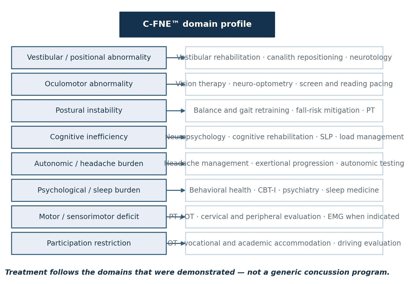
guide domain-specific referral and rehabilitation, so that treatment follows the systems actually demonstrated to be abnormal;
support serial comparison, so that change over time is assessed against a documented prior examination performed the same way;
provide the examination foundation for the Short-Term Concussion Disability (ST-CD) Scale, which converts selected C-FNE findings into Functional System grades and a single current disability score.21
The relationship in that final item is directional and asymmetric, and is developed in detail in Sections 3.5 and 16.0. The C-FNE can be performed, reported, and clinically used without ever generating an ST-CD score. The ST-CD score cannot be generated, reported, or interpreted without the underlying C-FNE data.
Terminology
The following definitions govern usage throughout this paper. Several are drawn from the World Health Organization International Classification of Functioning, Disability and Health (ICF), which supplies the impairment–activity–participation architecture used here.
Table 1. C-FNE terminology and definitions.
Term
Definition as used in this paper
Concussion-type traumatic brain injury (cTBI)
A descriptive term for the clinical entity commonly labeled concussion or mild traumatic brain injury: a traumatically induced disturbance of brain function, with or without loss of consciousness, without a structural lesion requiring surgical management. Descriptive only; not a new diagnostic category.
C-FNE
The Comprehensive Functional Neurological Evaluation — the standardized, sequenced, multidomain examination framework described in this paper, comprising instrument-derived, clinician-administered, patient-reported, and participation-level components with defined quality controls and documentation requirements.
Functional System (FS)
One of the nine neurological or functional domains into which C-FNE findings are organized. The term is used in the sense established by ordinal neurological disability scaling, in which the profile of system-level involvement is retained alongside any global score.
Instrument-derived finding
A numerical result generated by a calibrated device under standardized conditions — for example slow-phase velocity in degrees per second, saccadic latency in milliseconds, pursuit gain, center-of-pressure excursion, or grip force in kilograms.
Standardized clinical finding
An observation made by a trained examiner following a defined procedure with defined normal and abnormal categories — for example a cranial nerve screening result or an observed nystagmus response to a positional maneuver.
Patient-reported outcome (PRO)
A measurement of a health state reported directly by the patient without interpretation by a clinician, obtained here through validated instruments such as the Insomnia Severity Index, PHQ-9, and GAD-7.
Impairment
A problem in body function or structure, such as a significant deviation or loss (ICF). In this framework: an abnormal measured or observed neurological finding.
Activity limitation
Difficulty an individual may have in executing activities (ICF). In this framework: demonstrated difficulty performing a discrete task such as reading, driving, or walking on uneven ground.
Participation restriction
A problem an individual may experience in involvement in life situations (ICF). In this framework: reduced or altered engagement in work, school, household, social, or community roles.
Short-term disability
The current, potentially recoverable functional consequence of impairment, measured at a defined point in the post-injury course before maximum medical improvement. This is what the ST-CD Scale quantifies.
Permanent impairment
A determination of residual, non-recoverable loss made after maximum medical improvement, under an established impairment-rating framework. The C-FNE and ST-CD do not establish permanent impairment.
The ICF definitions of impairment, activity limitation, and participation restriction are used as published.22 The distinction between short-term disability and permanent impairment follows the conventional separation between a recoverable clinical state and a post-maximum-medical-improvement rating.23
Three time-based terms are used repeatedly in this paper and are defined here so that they carry a consistent meaning throughout. For the purposes of this framework, the acute phase is the period from injury through fourteen days; the subacute phase runs from fifteen days through three months; and the chronic phase begins after three months. These boundaries are administrative conventions adopted for consistency of reporting, not biological thresholds, and they are stated explicitly because the interval since injury materially affects how C-FNE findings should be interpreted. The framework was developed for, and its interpretive rules are written for, the acute and subacute periods. Application in the chronic phase is discussed in Section 22.0.
Scope and intended audience
This white paper is a methodology document. It describes what the C-FNE examination consists of, how it is administered, how its outputs are reported, what interpretive rules govern it, and what it does and does not support. It is not a scoring manual, not a validation report, and not a clinical practice guideline.
It is written for the following readers:
neurologists and neurosurgeons managing post-traumatic patients;
physiatrists and rehabilitation physicians;
vestibular clinicians, audiologists, and neurotologists;
primary care and urgent care clinicians who receive these patients first;
physical, occupational, and speech-language therapists;
neuropsychologists and clinical psychologists;
behavioral health professionals;
attorneys and other medicolegal professionals evaluating functional neurological documentation;
insurers, claims professionals, and case managers;
clinical researchers in concussion, vestibular science, and outcome measurement.
Readers from the last three groups are asked to attend particularly to Sections 16.4, 21.0, and 22.0, which state in explicit terms the uses this framework does not support.
Population, scope, and exclusions
The framework is specified for a defined population, and applying it outside that population without qualification is a misuse of it. The specification below governs.
Table 2. Target population, scope of application, and exclusions.
Category
Specification
Primary population
Adults aged eighteen years and older evaluated after suspected or clinically diagnosed cTBI
Typical timing
The acute and subacute periods as defined in Section 1.4 — that is, up to three months after injury
Chronic application
Permitted, with explicit qualification that the framework’s interpretive rules were developed for earlier phases and that findings characterize present function rather than the original injury
Adolescents and children
Not specified by this framework. See the constraints below; adult reference ranges are not applied to minors without age-appropriate norms
Emergency or medical instability
Not appropriate. The examination is deferred until the patient is medically stabilized
Major neurological red flags
Requires urgent conventional medical evaluation before any C-FNE testing (Section 1.7)
Severe cognitive incapacity
Partial examination or deferral, with the affected components recorded as not tested and the reason stated
Uncorrected major sensory impairment
Testing modified, deferred, or qualified. Visual and auditory correction status is recorded, because several components depend on it
Suspected cervical instability
Positional and provocative vestibular testing withheld pending clearance
The C-FNE was developed in an adult outpatient population. Its testing sequence, its interpretive rules, and the reference ranges applied to its instrumented components were established in patients aged eighteen years and older, and the framework as described in this paper is specified for that population.
The framework is nonetheless administered in practice to adolescent patients, and that requires an explicit statement rather than silence. Several of the constraints below apply, and each is a limitation of the present framework rather than a solved problem:
the instrumented components — posturography, videonystagmography, oculomotor recording, and dynamometry — have age-dependent normative distributions, and the adult reference ranges used by this framework should not be applied to a minor without age-appropriate norms for the specific device and protocol in use;
the computerized cognitive platform and the psychological and sleep instruments carry their own separately published age ranges, and each must be used only within the range for which it is validated;
developmentally appropriate suicide-risk screening and escalation procedures differ for minors, including the circumstances under which a guardian is informed, and the escalation pathway in Section 9.5 must be adapted accordingly;
guardian consent and, where the patient is capable of it, patient assent are obtained before examination, and school and sport participation are documented alongside the work and driving items used for adults;
parent- or guardian-reported information about function is recorded, but it is recorded as a distinct information source and is not merged with the patient’s own report.
Until an explicit pediatric protocol with age-stratified reference data has been developed and published, the framework should be described as specified for adults. Where it is applied to an adolescent, the report should state the patient’s age, identify every reference range that was derived from an adult sample, and qualify each affected conclusion accordingly. Extension of the framework to a pediatric population is listed as a development priority in Section 23.0.
Red flags requiring evaluation before examination
This paper may be read by clinicians who are not neurologists, and the point therefore requires stating without hedging. The C-FNE is an elective outpatient examination of function. It is not a triage instrument, it is not sensitive to acute intracranial emergencies, and it is never a substitute for urgent conventional evaluation. Where any of the following is present, the examination is deferred and the patient is referred for urgent medical assessment:
declining or fluctuating level of consciousness;
new focal weakness, sensory loss, or asymmetry of the pupils;
repeated vomiting;
seizure activity, whether witnessed or reported;
severe or rapidly worsening headache, particularly of abrupt onset;
any clinical suspicion of intracranial hemorrhage or of a mass lesion;
acute visual loss or new diplopia;
unstable vital signs;
suspected cervical spine instability or fracture;
suicidal ideation with intent, plan, or means, which requires emergency intervention under the escalation pathway in Section 9.5 rather than continuation of testing.
The presence of a red flag is recorded, together with the referral made and its outcome. Where the patient later returns for examination, the record notes that the red flag was evaluated and addressed before testing proceeded. A C-FNE performed despite an unevaluated red flag is not a defensible examination, whatever its findings.
Origin and Development of the C-FNE™
Development history
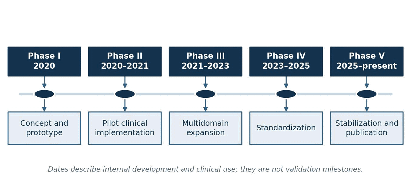
The C-FNE framework was conceived in approximately 2020 and has been in continuous clinical use and iterative refinement since. Its development was practice-driven rather than grant-funded: the structure emerged from the repeated experience of evaluating post-traumatic patients whose imaging was normal, whose symptom inventories were high but nonspecific, and whose available objective testing was scattered across several reports that no single clinician had integrated.
Five development phases are distinguished. The dates below describe internal development and clinical implementation. They are not validation milestones, and no phase should be read as establishing a measurement property.
Figure 1. C-FNE development phases. Dates describe internal development and clinical use, not validation.
Table 3. Development phases from 2020 to present.
Phase
Period
Principal developments
Phase I
2020
Concept and prototype. Clinical need formally identified. Initial examination components assembled from existing validated instruments. Early global clinical interpretation developed as an unstructured impression.
Phase II
2020–2021
Pilot clinical implementation. Balance, cognition, vestibular, and oculomotor testing organized into a single encounter. Examination sequence refined to manage symptom provocation and fatigue. Initial reporting templates developed.
Phase III
2021–2023
Multidomain expansion. Psychological and sleep measures added. Cranial nerve and sensorimotor screening formalized. Quantitative strength testing and functional participation measures incorporated. Cross-domain interpretation rules developed.
Phase IV
2023–2025
Standardization. Testing sequence standardized. Abnormality thresholds formalized against device and published reference ranges. Incomplete-test and technical-failure rules developed. Quality-control procedures refined. Relationship to the ST-CD Scale defined.
Phase V
2025–present
Stabilization and publication. Protocol placed under version control. Examination architecture stabilized. White paper and peer-reviewed methodology manuscripts prepared. Formal validation program planned (Section 23.0).
Development setting
The framework was developed in an outpatient multidisciplinary practice providing neurological, vestibular, rehabilitative, and behavioral health services across several states. The patient population consists predominantly of adults and adolescents in the acute and subacute period following cTBI, typically evaluated between several days and several months after injury, with a smaller number evaluated in the chronic phase.
Common mechanisms in the development population include motor vehicle collisions, falls, occupational injuries, assaults, and sport-related injuries, with motor vehicle collision the single most frequent. This distribution matters, because it differs materially from the populations in which most concussion assessment tools were validated.
That difference explains why existing emergency-department and sideline instruments were not sufficient for this setting. Sideline and acute-assessment tools are designed for rapid screening of a witnessed injury in a known individual, often with a pre-injury baseline available, minutes to hours after the event.24–26 Their design constraints are speed, portability, and immediate return-to-play decision support. The population addressed by the C-FNE arrives days to months later, usually without a baseline, frequently with polytrauma, pain, medication effects, and psychological sequelae superimposed, and requires a treatment plan rather than a removal-from-play decision. Emergency-department instruments, similarly, are constructed to identify patients needing neuroimaging or admission and are explicitly not designed to characterize functional impairment. Neither category of tool answers the question that dominates outpatient management: which neurological systems are impaired, to what degree, and what should be done about each of them.
Development objectives
The C-FNE was designed against eight explicit objectives. Each carries a corresponding cost, and those costs are stated because a framework that presents only its advantages is not a serious methodological document.
Table 4. Development objectives and their accepted costs.
Objective
What it requires
Accepted cost
Multidomain
Nine domains examined in every complete evaluation rather than testing only the presenting complaint
Longer encounter; higher patient burden; some domains will be normal in most patients
Standardized
Fixed sequence, fixed conditions, fixed reporting format
Reduced flexibility to adapt the examination to an individual presentation
Clinically feasible
Deliverable in a single outpatient session with existing commercially available equipment
Exclusion of research-grade methods (e.g., rotary chair, VEMP, formal neuropsychological batteries) from the core protocol
Reproducible in procedure
Written administration rules, examiner training, documented deviations
Procedural reproducibility is not the same as measurement reliability, which remains unestablished (Section 22.0)
Transparent
Every conclusion traceable to a stated measurement and reference range
Longer reports; visible disclosure of incomplete and equivocal findings
Auditable
Preservation of raw values, device identity, software version, and examiner identity
Substantially greater documentation burden
Serially administrable
Identical procedure at each visit; prior results available at interpretation
Repeated administration carries practice and habituation effects, particularly for cognitive and positional testing
Cross-specialty intelligible
Common vocabulary; component results reported in their native units alongside interpretation
Some loss of discipline-specific nuance in the summary layer
Table 5. Design-control matrix: clinical problem, design response, and current validation status.
Clinical problem
Design requirement
Implemented feature
Verification method
Validation status
Testing fragmented across settings and dates
One sequenced examination in a single encounter
Nine-domain protocol, fixed order, single session
Protocol audit against the written sequence
Not yet validated
Missing data silently read as normal
Explicit completion categories that cannot default to normal
Not indicated / not tested / aborted / invalid / uninterpretable labels (Table 31)
Report audit for unlabelled omissions
Planned — Phase 2
Fatigue and provocation contaminating later components
Standardized test order
Cognitive and oculomotor testing before provocative vestibular procedures
Protocol audit; deviation log review
Planned — Phase 2
Conclusions not traceable to data
Every conclusion referable to a measured value
Raw values, units, and reference ranges preserved in the report
Independent reconstruction of domain conclusions
Planned — Phase 2
Device summary flags disagreeing with the underlying record
Data govern over device-generated flags
Interpretation from numerical values and tracings; discrepancies documented
Blinded re-read of recorded studies
Planned — Phase 2
Subjective measures presented as physiological tests
Information sources kept separate and labelled
Four-source model with per-source evidentiary weight (Table 6)
Report audit for source labelling
Planned — Phase 1
Serial comparison without a measurement-error estimate
Change interpreted only against measurement error
Serial-testing rules (Section 18.6); change reporting deferred pending data
Test–retest study in a stable subgroup
Planned — Phase 3
Examination outputs used beyond their evidentiary reach
Explicit statement of unsupported uses
Boundary statements in Sections 3.1, 15.4, 16.4, and 21.0
Medicolegal review of issued reports
Planned — Phase 1
The right-hand column is the operative one. Every entry in it is either “not yet validated” or “planned,” and that is the accurate present state of the framework. A design requirement that has been implemented is not thereby validated; implementation is a claim about what the procedure does, and validation is a claim about how well it does it. The two are separated here deliberately, and they remain separated until the studies in Section 23.0 are complete.
Trademark and authorship statement
The term Comprehensive Functional Neurological Evaluation and the abbreviation C-FNE were introduced by Wayne Croft during development of the examination framework. C-FNE is used to identify the standardized multidomain evaluation described in this white paper. Trademark designation identifies the framework and does not imply that the individual component tests are proprietary to the developer.
This qualification is material rather than formal. The C-FNE incorporates independently developed, separately published, and in several cases commercially licensed instruments — among them videonystagmography systems, force-platform posturography, computerized cognitive testing platforms, hand dynamometry, and validated patientreported instruments including the Insomnia Severity Index, PHQ-9, GAD-7, a PTSD screening measure, and the Columbia-Suicide Severity Rating Scale. Those instruments belong to their respective developers, retain their own validation literature, their own reference ranges, and their own scoring rules, and are used here as published. What the trademark identifies is the assembly, sequencing, quality control, documentation structure, and interpretive framework — not the components.
Section 21.4 addresses the further and equally important point that trademark ownership is a commercial designation and carries no scientific weight whatever: it does not validate the framework, establish accuracy, or substitute for independent replication.
Conceptual Foundation
The examination is not the diagnosis
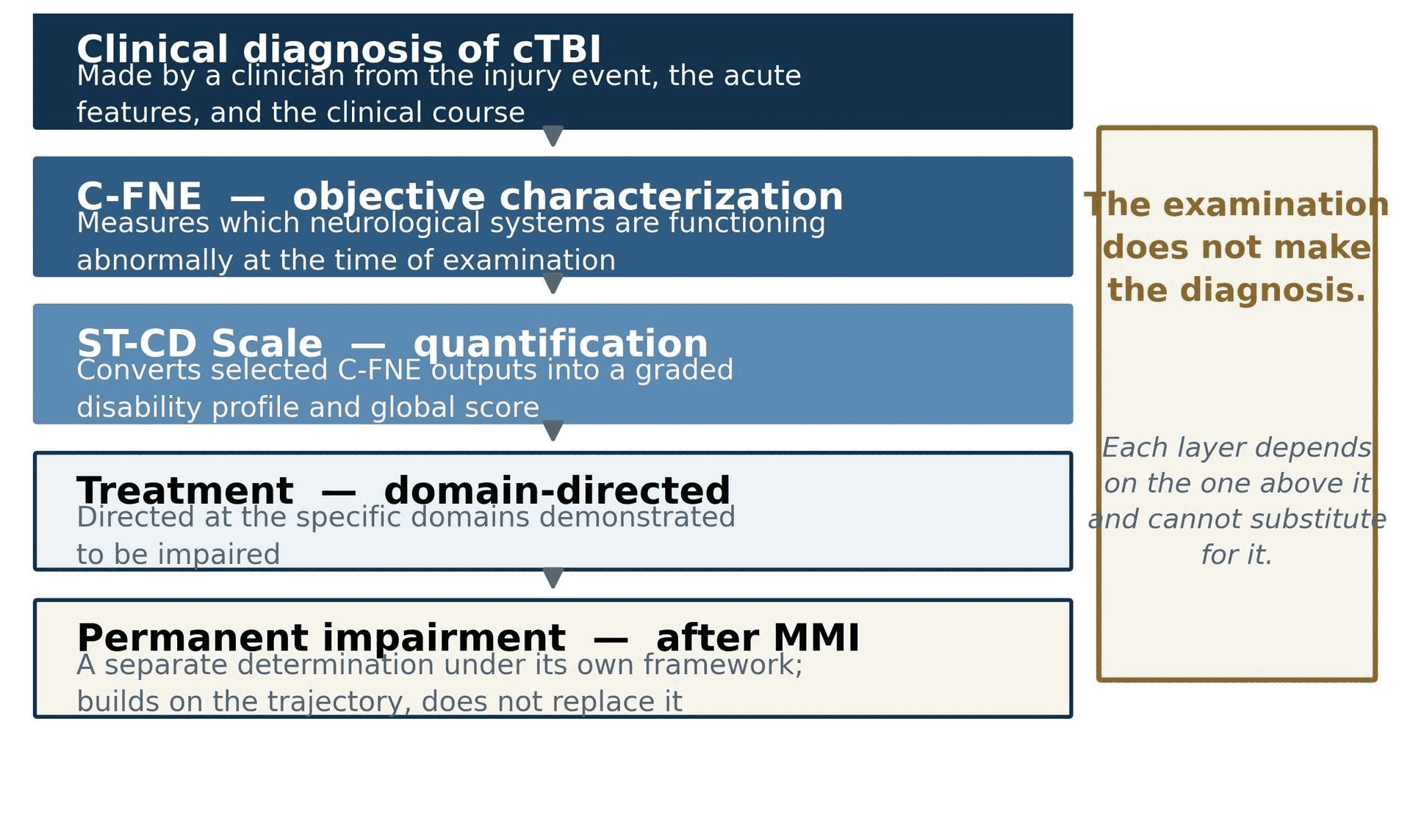
The diagnosis of cTBI is clinical and is made from the injury history, mechanism, acute signs and symptoms, medical records, examination findings, and differential diagnosis. The C-FNE™ does not independently determine whether a concussion occurred. It identifies and characterizes current functional abnormalities that may be relevant to the clinical evaluation.
This is the single most important boundary in the framework, and it is stated first because every subsequent interpretive rule depends on it. No pattern of C-FNE findings — however extensive, however internally coherent — constitutes a diagnosis of concussion. The diagnostic criteria for cTBI are event-anchored: they require a plausible mechanism of biomechanical force transmitted to the head or body, together with acute clinical signs occurring in temporal association with that event.27,28 Those criteria are met or not met by the history and the contemporaneous record. An examination performed weeks later measures the patient’s current functional state; it does not observe the injury.
The corollary is equally important and less often stated: a completely normal C-FNE does not exclude a cTBI. Patients recover. Patients compensate. Some deficits are intermittent, load-dependent, or provoked only by conditions the examination does not replicate. A normal examination performed at one time point establishes that the measured domains were within reference limits at that time point, and nothing more.
Figure 2. The diagnostic hierarchy. Diagnosis is clinical and event-anchored; the C-FNE characterizes current function; the ST-CD Scale quantifies it. Each layer depends on the one above and cannot substitute for it.
Function rather than structural pathology
Three distinct kinds of information are commonly conflated in the assessment of this population, and separating them clarifies what the C-FNE does.
Structural imaging asks whether tissue is anatomically abnormal. It answers questions about hemorrhage, contusion, mass effect, shear injury visible at the resolution of the sequence, and structural lesions requiring intervention. It is the correct test for those questions and a poor test for functional ones. As noted in Section 1.1, it is normal in most patients with persistent post-concussive impairment.9,11
Bedside neurological examination asks whether the nervous system is grossly intact. Cranial nerves, strength, tone, reflexes, coordination, and sensation are assessed against categorical norms designed to detect focal lesions. This examination is highly sensitive to stroke, mass lesion, and peripheral nerve injury, and comparatively insensitive to the graded performance decrements characteristic of cTBI. A patient with disabling visual-motion intolerance, a pursuit gain well below reference, and a fall-risk-level posturography score can have an entirely normal bedside neurological examination.
Functional performance measurement asks how well a neural system performs a defined task under standardized conditions, and expresses the answer as a number on a continuous or ordinal scale against a reference distribution. Saccadic latency in milliseconds, pursuit gain as a ratio, slow-phase velocity in degrees per second, center-ofpressure excursion, reaction time, grip force — these are graded measures of process. This is the level at which cTBI abnormality predominantly resides, and it is the level at which the C-FNE operates.
None of the three replaces the others. Structural imaging remains mandatory where a surgical or progressive lesion is possible; the bedside examination remains the front line for focal neurological disease; and functional measurement, which is the subject of this paper, characterizes the graded process abnormalities that the other two are not designed to detect.
Multimethod assessment
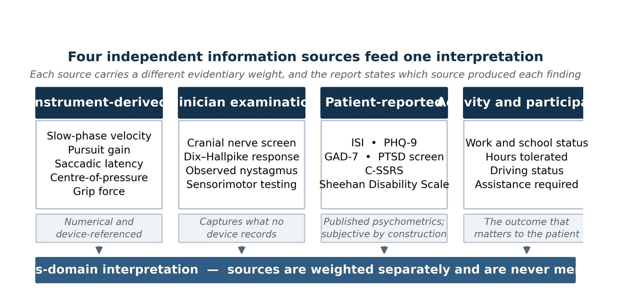
C-FNE information is drawn from four sources with materially different evidentiary properties. The framework requires that these be labeled and kept distinct, because treating them as interchangeable is a common and consequential error.
Figure 3. The four information sources feeding cross-domain interpretation. Each retains its own evidentiary weight.
Table 6. The four C-FNE information sources and their properties.
Source
Examples
Strength
Limitation
Instrument-derived measurement
VNG slow-phase velocity, saccadic latency and accuracy, pursuit gain, center-of-pressure excursion, computerized reaction time, dynamometric grip force
Numerical, device-referenced, minimally dependent on examiner judgment, reproducible in units
Sensitive to calibration, artifact, effort, medication, fatigue; abnormal values are rarely specific to cTBI
Standardized clinician examination
Cranial nerve screening, positional and Dix–Hallpike responses, observed nystagmus direction and latency, sensorimotor testing
Captures findings no device records; permits real-time judgment about validity and tolerance
Examiner-dependent; interrater agreement not yet established for this protocol
Published psychometrics; measures internal states no instrument can access; directly relevant to function
Subjective by construction; influenced by mood, expectation, context, and external incentive; never described as objective physiological tests
Documented activity and participation limitation
Work and school status, hours tolerated, accommodations, missed days, driving status, assistance required
Measures the outcome that actually matters to the patient; anchors severity to real-world consequence
Heavily conditioned by job demands, family support, economic circumstance, accommodation availability, and compensation context
The reporting rule that follows is simple and is enforced throughout: every finding in a C-FNE report must be identifiable as belonging to one of these four categories. A patient-reported score is never presented as an objective test result, and an instrument value is never presented as a symptom.
Cross-domain triangulation
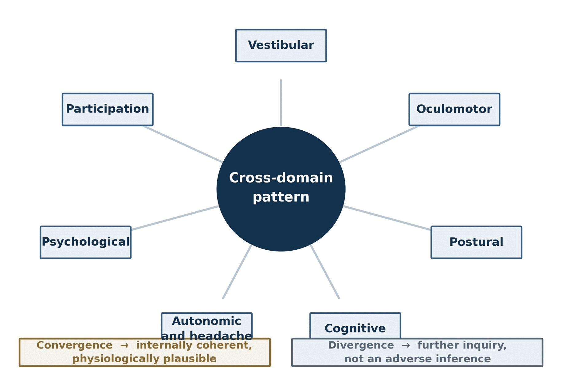
The interpretive value of a multidomain examination lies substantially in the relationships among domains rather than in any single result. Confidence that a finding reflects genuine physiological dysfunction increases when findings across independent domains are physiologically coherent — that is, when they are the pattern one would predict if the underlying dysfunction were real.
Examples of physiologically coherent cross-domain patterns include:
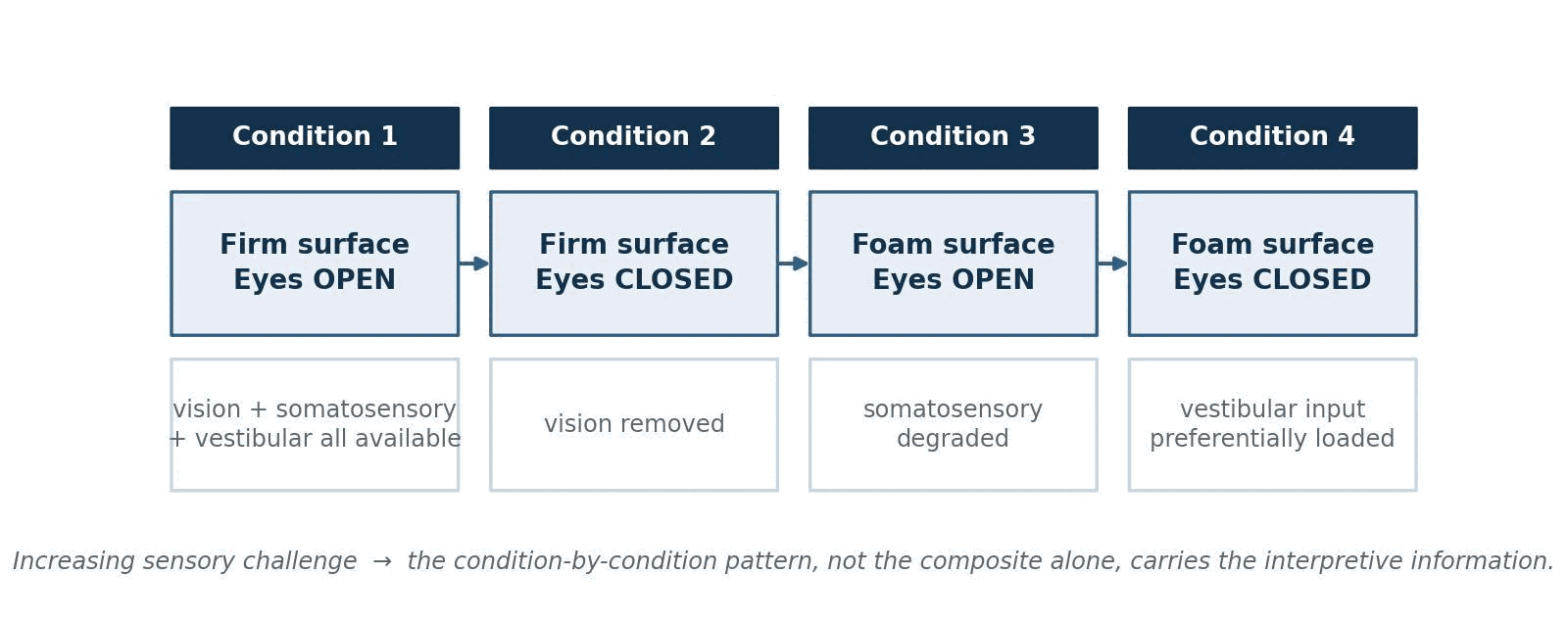
vestibular abnormality accompanied by impaired postural control, particularly with disproportionate degradation in the eyes-closed foam condition where vestibular input is preferentially loaded;29,30
oculomotor abnormality accompanied by documented intolerance of visual tasks — reading, screen work, scrolling, driving in visually complex environments;31,32
insomnia and fatigue accompanied by reduced cognitive efficiency, with the pattern of cognitive decrement weighted toward sustained attention and processing speed rather than storage;33
motor asymmetry on dynamometry accompanied by documented functional difficulty using that limb, and by a consistent sensory or reflex correlate.
Two cautions are essential and are stated as a formal rule of the framework.
Cross-domain convergence does not prove causation or effort, and divergence does not establish exaggeration or absence of injury.
Convergent findings are consistent with a real, physiologically organized dysfunction; they do not identify what caused it, and they do not exclude a pre-existing or unrelated condition producing the same coherent pattern. Divergent findings — a patient with severe reported dizziness and a normal vestibular battery, or normal reported function with markedly abnormal posturography — are common, have many innocent explanations (intermittent symptoms, compensation, testing on a good day, provocation thresholds not reached, medication, anxiety, pain), and are not evidence of exaggeration. Determinations regarding effort and response validity require dedicated performance-validity and symptom-validity methodology, applied and interpreted by qualified examiners, and are outside the scope of this framework.34–36
Figure 4. The cross-domain triangulation model. Convergence may increase confidence that the observed pattern is internally coherent and physiologically plausible; divergence prompts further inquiry rather than an adverse inference.
Relationship to the ST-CD Scale
The C-FNE and the ST-CD Scale are distinct instruments with distinct functions. The C-FNE is a procedure: it specifies what is measured and how. The ST-CD Scale is a scoring model: it specifies how a defined subset of those measurements, together with functional anchors, is converted into ordinal Functional System grades and a single global score. The relationship is deliberately one-directional.
Table 7. C-FNE versus ST-CD Scale.
C-FNE
ST-CD Scale
Standardized examination framework
Ordinal disability scoring framework
Produces component findings
Organizes selected findings into Functional System grades
Identifies which systems are abnormal
Characterizes overall current disability
Can be interpreted without a global score
Cannot be interpreted without supporting C-FNE data
Requires technical and clinical interpretation
Requires scoring and anchor reconciliation
Answers: which systems, how abnormal, measured how
Answers: how disabled is this patient right now
A procedure
A scoring model applied to the output of that procedure
It is worth noting what the ST-CD is not intended to duplicate. Established outcome and disability instruments — the Extended Glasgow Outcome Scale, the Functional Independence Measure, and comparable tools — were designed for populations and severity ranges in which the discriminating questions differ substantially from those posed by an ambulatory, independently functioning patient with distributed functional impairment.37,38 Their floors and ceilings sit in the wrong place for this population, which is the gap the ST-CD was constructed to address.
The analogy the developers have used internally is that the C-FNE is the engine and the ST-CD is the speedometer. The speedometer reports a single number that is genuinely useful and genuinely informative, and it is meaningless without the machinery that produces the motion. A speedometer reading reported without any indication of what vehicle it came from, whether all cylinders were firing, or whether the instrument was calibrated is not a measurement — it is a number. This is the reasoning behind the reporting rule in Section 16.3, which prohibits reporting an ST-CD global score without the underlying Functional System profile and component findings.
Relationship to existing assessment instruments
The C-FNE is assembled from independently developed instruments and sits alongside established assessment approaches rather than replacing them. Because a framework presented by its own developers invites the suspicion that it has been positioned as superior to everything else, the comparison below is stated in terms of design intent. Each instrument listed does something the others do not, and in several rows the honest entry in the final column is that the C-FNE adds nothing.
Table 8. The C-FNE in relation to established assessment approaches. Each approach is designed for a different question; none is a general substitute for another.
Instrument or approach
What it is designed to do
Scope
What the C-FNE adds in this setting
Routine neurological examination
Detect focal neurological disease and localize structural lesions
Categorical, bedside, no instrumentation
Graded quantitative measurement of process abnormalities that a categorical bedside examination is not designed to detect
SCAT and equivalent sideline tools
Rapid screening of a witnessed injury within minutes to hours, often against a pre-injury baseline
Acute, brief, screening
Nothing in the acute sideline setting, where these tools are the appropriate instrument. The C-FNE addresses the later outpatient encounter, where no baseline exists
VOMS
Screen for symptom provocation by vestibular and ocular motor tasks
Symptom-provocation screening, clinicianobserved
Instrumented measurement of the same systems in physical units, alongside the other seven domains
BESS
Screen postural stability without instrumentation
Observational balance scoring
Instrumented four-condition posturography with a sensorycondition profile rather than an error count
Computerized cognitive testing alone
Quantify cognitive efficiency against normative data
Single domain
Interpretation of the same scores in the context of sleep, mood, pain, and vestibular findings measured at the same encounter
Formal neuropsychological evaluation
Comprehensive characterization of cognition with validity testing
Single domain, in depth; several hours
Nothing in depth of cognitive assessment, where formal evaluation is superior. The C-FNE screens cognition and refers to formal evaluation when indicated
Isolated VNG
Characterize vestibular and oculomotor function
One or two domains; reported to the ordering clinician
Integration of the VNG result with balance, cognitive, autonomic, psychological, and participation findings in one report
Comprehensive vestibular laboratory testing
Full characterization of vestibular function including rotary chair, VEMP, and vHIT
One domain, exhaustively
Nothing in vestibular depth, where the laboratory is superior. The C-FNE is a clinically deliverable battery that refers onward when the vestibular question exceeds its resolution
Three of these rows deserve emphasis because they run against the interest of the framework’s developers. In the acute sideline setting the sideline tools are the correct instrument and the C-FNE has no role. Where the question is the depth and validity of cognitive impairment, formal neuropsychological evaluation is superior and the C-FNE refers to it. Where the question is the precise characterization of vestibular physiology, a comprehensive vestibular laboratory is superior and the C-FNE refers to it. The framework’s contribution is breadth in a single encounter with preserved component detail, and breadth is bought at the cost of depth in every individual domain.
C-FNE™ Examination Architecture
Overview of the testing process
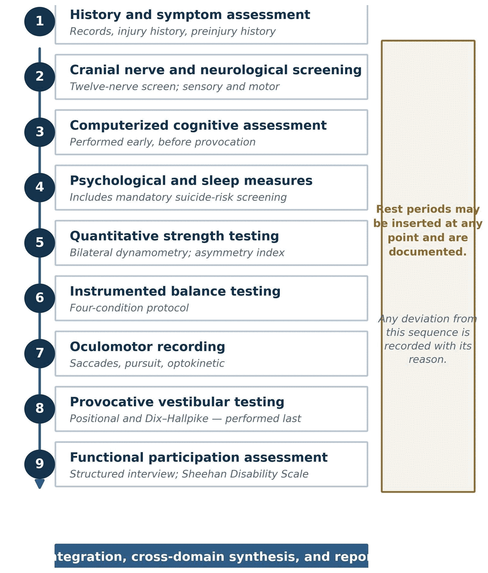
A complete C-FNE proceeds through a defined sequence of ten stages. The sequence is not arbitrary; it reflects deliberate decisions about symptom provocation, fatigue, calibration dependency, and the order in which information becomes available to the interpreting clinician.
Figure 5. The C-FNE examination workflow. Rest periods may be inserted at any point; every deviation from the standard sequence is documented.
Stage one, referral and records review, occurs before the patient is seen. Prior imaging, emergency-department records, prior concussion history, current medications, and any previous vestibular, cognitive, or psychological testing are reviewed, because each of these conditions the interpretation of what follows. An examination interpreted without knowledge that the patient takes a vestibular suppressant, or had a prior vestibular diagnosis, or has an established learning disability, is an examination interpreted incorrectly.
The final stage — recommendations and, when applicable, ST-CD scoring — is deliberately last and deliberately conditional. The examination is complete and reportable whether or not a disability score is generated.
Examination duration
A complete C-FNE generally requires approximately 90 to 150 minutes, depending on symptom tolerance, the need for rest periods, calibration difficulty, and whether all testing components are clinically indicated.
That range is wide because the principal determinant of duration is the patient, not the protocol. Patients with significant visual-motion intolerance, headache, or anxiety frequently require multiple rest periods and occasionally cannot complete the vestibular portion in a single session. Calibration of the video-oculography system can be prolonged or unsuccessful in patients with ptosis, heavy eye makeup, corrective lenses, marked photophobia, or difficulty maintaining fixation. Where duration is extended by these factors, the reason is recorded, because prolonged testing is itself a fatigue confounder for whatever is tested last.
Where the full examination cannot be completed in one session, the protocol permits division across two sessions within a short interval, with the date of each component recorded separately. It does not permit silently omitting components, and it does not permit reporting a partial examination as a complete one.
Standard testing sequence
The order of testing is a substantive methodological decision rather than a scheduling convenience. Four principles govern it.
Cognition is tested before provocative vestibular procedures. Positional testing, caloric irrigation, and optokinetic stimulation reliably provoke dizziness, nausea, headache, and autonomic symptoms in this population. Cognitive testing performed afterward measures cognition under provoked symptom load, which is not the intended construct and is not comparable to the normative sample. Cognitive testing is therefore placed early, before the patient has been made symptomatic by the examination itself.
Provocative positional and caloric testing are placed late. The same reasoning applies in reverse: these procedures are the most likely to end the session, and placing them last ensures that the maximum number of domains has already been recorded before tolerance becomes the limiting factor. Caloric irrigation in particular is the least welltolerated component of the standard battery and is positioned at or near the end.39
Rest periods are permitted and documented. A patient who requires a fifteen-minute recovery period between positional testing and strength testing should be given it, and the interruption should appear in the record, because the results after a long rest are not directly comparable to results obtained under continuous load.
Deviations from sequence are documented. Where the order is altered — for equipment availability, patient tolerance, scheduling, or clinical judgment — the deviation and its reason are recorded. This is a specific requirement rather than a general aspiration, because sequence effects are a plausible source of variance and cannot be evaluated retrospectively if they were not recorded.
Core versus conditional components
The C-FNE distinguishes core components, which are performed in every complete examination, from conditional components, which are performed when specifically indicated. This distinction protects the interpretability of the examination: if the composition of the battery varied freely from patient to patient, no serial or cross-patient comparison would be possible.
A distinction that is easily misread should be stated directly. Every complete C-FNE evaluates all nine functional domains. A domain may be evaluated through its core procedures alone, or through those core procedures supplemented by conditional testing. Conditional testing expands the characterization of a domain; it does not create the domain, and its absence does not leave the domain unexamined. Calorics, orthostatic testing, exertional tolerance testing, and supplemental neuropsychological testing are therefore not mandatory in every case, and an examination that omits them where they were not indicated is a complete examination rather than a partial one. What is never permissible is the reverse: omitting a core component and reporting the examination as complete. The rules distinguishing those two situations are set out in Section 4.6.
Table 9. Core and conditional examination components.
Component
Status
Indication or note
History and symptom assessment
Core
Performed in all cases; anchors every subsequent interpretation
Cranial nerve and neurological screening
Core
Screening level; not a substitute for a complete neurological examination
Instrumented balance testing
Core
Four-condition protocol; deferred only for orthopedic or safety contraindication
Computerized cognitive testing
Core
Performed early in the sequence, before provocative testing
Structured interview plus validated participation measure
Caloric irrigation
Conditional
Indicated where unilateral or bilateral peripheral hypofunction is suspected; contraindicated with tympanic membrane perforation or active ear disease
Orthostatic testing
Conditional
Indicated for reported positional lightheadedness, syncope, or autonomic symptoms
Exertional tolerance testing
Conditional
Indicated for return-to-activity planning and physiological subtype clarification; requires cardiovascular clearance
Additional neuro-ophthalmic examination
Conditional
Indicated for convergence insufficiency, diplopia, accommodative dysfunction, or visual field concern
Supplemental neuropsychological testing
Conditional
Indicated where computerized screening is abnormal, invalid, or discrepant with function
Advanced imaging recommendation
Conditional
Indicated for focal deficit, red-flag features, atypical course, or suspicion of alternative pathology
Specialist referral
Conditional
Directed by the demonstrated domain profile (Section 17.1)
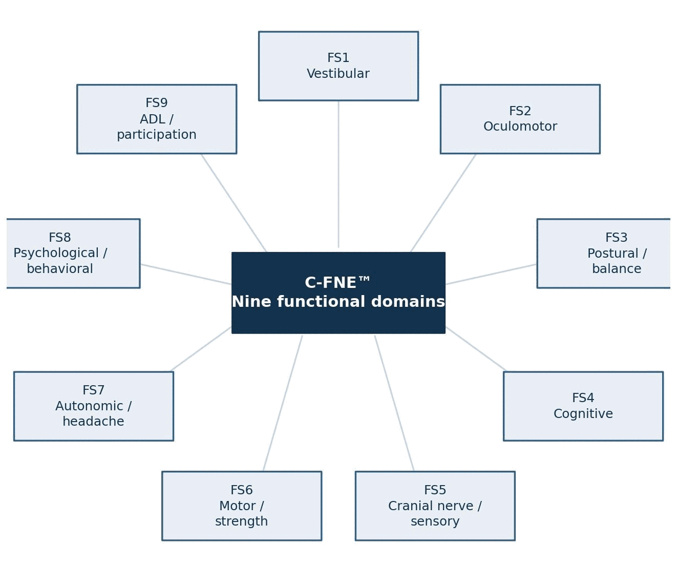
The nine functional domains into which findings are organized are set out below. These domains structure the report, drive the referral logic in Section 17.1, and correspond to the Functional Systems used by the ST-CD Scale.
Figure 6. The nine C-FNE functional domains.
Table 10. Nine functional domains and their principal measures.
Table 11. Mapping of the eight core examination components to the nine functional domains.
Core examination component
Functional domain(s) informed
Note
History and symptom assessment
Contextualizes all nine (FS1–FS9)
Anchors interpretation; not itself a graded domain
Cranial nerve and neurological screening
FS5 cranial nerve and sensory
Also contributes observations relevant to FS2 and FS6
Instrumented balance testing (fourcondition)
FS3 postural control
Sensory-condition pattern also informs FS1
Computerized cognitive testing
FS4 cognitive
Performance is influenced by FS7 and FS8; interpreted jointly
Psychological and sleep screening
FS8 psychological and behavioral
Sleep and mood findings materially affect FS4 interpretation
Quantitative strength assessment
FS6 motor and strength
Asymmetry index is the principal output
Videonystagmography and oculomotor testing
FS1 vestibular and FS2 oculomotor
One recording session yields two separately graded domains
Functional participation assessment
FS9 activities of daily living and participation
Also supplies the headache and autonomic history informing FS7
One inference the reader should not draw is that nine domains imply nine equal domains. The C-FNE itself applies no weighting. It is an examination: it measures each domain, reports each result in its own units, and states a conclusion for each. It does not rank the domains against one another, and it does not combine them into any single quantity. Clinically the domains are plainly not of equal consequence to every patient — a severe balance deficit and a mild oculomotor finding have very different implications for safety and for return to work — but that judgment is made by the clinician against the individual’s circumstances, not encoded in the examination. Weighting enters only when STCD scoring is applied, and it enters there through the scale’s own derivation rules, which are stated in the ST-CD specification rather than in this paper. A reader who wishes to know how the domains are combined should consult that specification; a reader who wishes to know what was measured should read the component results.
The counts differ because the relationship is not one-to-one. A single recording session yields both the vestibular and the oculomotor domain; the headache and autonomic domain draws on history taken during two different components; and history and symptom assessment informs the interpretation of every domain without being graded as one. The nine domains are the reporting and grading structure; the eight core components are the procedures performed to populate them.
Verification note. The nine-domain structure above is presented as the framework’s architecture and must be reconciled against the developer’s authoritative source documents — the C-FNE report workbook and the ST-CD scoring specification — before external publication. Per-domain numeric abnormality thresholds and normative reference tables are deliberately not stated in this white paper; they are protocol-level and device-level parameters that belong in the administration manual (Supplement A) where they can be version-controlled independently of this document.
The core vestibular battery and the role of caloric testing
Because caloric irrigation is classified as conditional rather than core, the framework must state what the vestibular domain can and cannot conclude when calorics are not performed. This is not a hypothetical concern: caloric testing is among the least well tolerated procedures in the battery, and it is aborted or declined in a substantial proportion of symptomatic patients.
The core vestibular battery consists of spontaneous nystagmus testing with and without fixation, gaze-evoked nystagmus testing in the cardinal positions, positional testing, and Dix–Hallpike testing, each recorded and reported with laterality, beat direction, and measured slow-phase velocity in every position tested. An examination containing all of these has a complete core vestibular battery, and the vestibular domain may be graded from it.
What calorics uniquely answer. Caloric irrigation is the only component of the battery that tests each horizontal semicircular canal independently at low frequency. It is therefore the only component that quantifies unilateral peripheral hypofunction — expressed as unilateral weakness and directional preponderance — and the only one capable of demonstrating bilateral vestibular hypofunction. No other component of the core battery substitutes for it. A patient with normal spontaneous, gaze, positional, and Dix–Hallpike findings may still have significant peripheral hypofunction that only calorics would reveal.
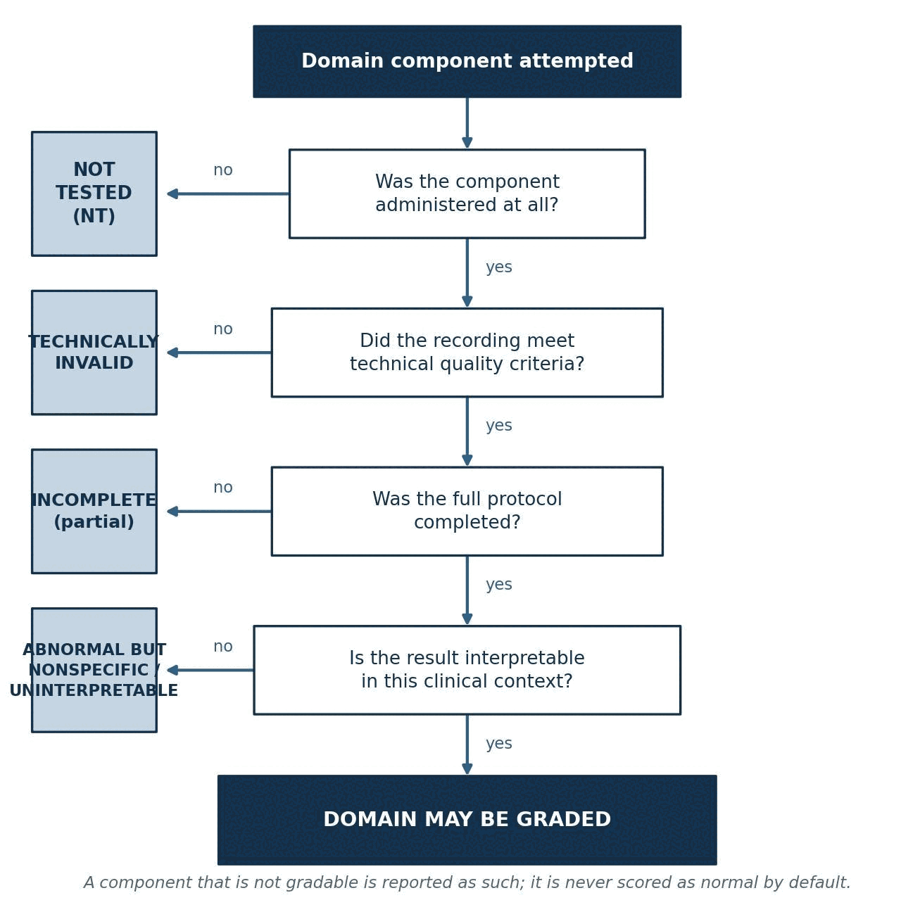
“Not indicated” is not “not tested.” Where the clinical picture gives no reason to suspect peripheral hypofunction and calorics were deliberately not ordered, the component is recorded as not indicated, and the examination remains complete. Where calorics were indicated and were attempted but not completed, the component is recorded as aborted, with the number of irrigations obtained and the reason for stopping. Where they were indicated and not attempted at all, the component is not tested. These three states carry different information and are never recorded interchangeably.
Consequences for interpretation. Where calorics are absent for any reason, the vestibular domain may still be graded on the core battery, but the report states explicitly that peripheral hypofunction has not been excluded, and no conclusion of “normal peripheral vestibular function” is issued. Where calorics were partially obtained — for example two of four irrigations — the individual responses obtained are reported as measured values, and the derived indices requiring the full protocol are not computed. An absent response on a partial battery cannot be distinguished from a technical failure and is reported as uninterpretable rather than as absent function.
Consequences for ST-CD scoring. Vestibular grading may proceed without caloric results provided the core battery was validly obtained. The grade is then assigned on the demonstrated findings, and the absence of caloric data is disclosed alongside the grade. A grade assigned without calorics is not marked down for the missing component, and neither is it marked up: the framework grades what was demonstrated, and discloses what was not examined.
Defining a complete examination
Section 19.0 states that a partial examination may not be represented as complete. That rule requires a minimum dataset, which is specified here.
Table 12. Minimum dataset for a complete C-FNE examination.
Required element
Complete
Partial
Not applicable
Records review and injury history
Required
Cannot be omitted — examination is not interpretable without it
—
Cranial nerve and neurological screening
Required
NT with reason if the patient cannot participate
—
Computerized cognitive assessment
Required
NT if unable, declined, deferred, or technically invalid
—
Four-condition instrumented balance testing
Required
NT if contraindicated or unsafe
—
Core VNG and oculomotor battery
Required
NT if not attempted; aborted or invalid if attempted and unusable
—
Quantitative strength testing
Required
NT if orthopedically contraindicated
—
Psychological and sleep measures
Required
NT if declined or invalid; suicide-risk screening is never omitted
—
Functional participation assessment
Required
Cannot be omitted — supplies the FS9 grade
—
Caloric irrigation
Where indicated
Aborted or NT as applicable (Section 4.5)
Not indicated where peripheral hypofunction is not suspected
Orthostatic, exertional, neuroophthalmic, and supplemental neuropsychological testing
Where indicated
Aborted or NT as applicable
Not indicated in the absence of the triggering finding
The three reporting labels follow directly from the table and are applied to the examination as a whole:
Complete C-FNE. Every core component was validly obtained, and every conditional component that was indicated was either obtained or explicitly recorded as aborted or not tested with a stated reason. All nine domains are gradable.
Partial C-FNE. One or more core components were not validly obtained. The report identifies which, states the reason, and grades only the domains for which the minimum component set was obtained. A partial examination remains clinically useful — it identifies the abnormalities that were demonstrated and directs treatment to them — and it is labelled partial on its face.
C-FNE attempted but not interpretable. So few components were validly obtained that no cross-domain synthesis is possible. The individual results obtained are reported as measured values, no domain conclusions and no global interpretation are issued, and re-examination is recommended with the specific barrier identified.
The label appears in the report header, not only in the body, so that a reader who sees only the first page cannot mistake a partial examination for a complete one. No ST-CD global score is issued from an examination in the third category, and a score issued from an examination in the second is accompanied by the list of ungraded domains.
Completeness is not one thing, and four senses of it are distinguished because they are routinely conflated. An examination can be clinically complete and protocol incomplete at the same time, and that combination is common rather than exceptional.
Table 13. Four senses of a complete examination.
Tier
Definition
Who relies on it
Clinically complete
Sufficient to answer the referral question and to direct treatment, whether or not every core component was obtained
Treating clinicians. A partial examination is frequently clinically complete
Protocol complete
All core components validly obtained, and every indicated conditional component either obtained or labelled with its reason (Table 12)
The framework itself. This is the tier the label “Complete C-FNE” denotes
ST-CD eligible
Contains the minimum valid data required for the scale’s grading rules, with any ungraded domains disclosed alongside the score
Anyone issuing or reading an ST-CD score
Research complete
Meets a study’s predefined dataset, device, timing, and quality criteria for inclusion in analysis
Investigators. Set by each study protocol, and stricter than protocol complete
The distinction matters most for anything retrospective. A dataset assembled by pulling every examination labelled complete will mix examinations performed on different devices, at different intervals after injury, and under different protocol versions. Those are not equivalent observations, and pooling them would produce a reliability or outcome estimate that is an artifact of the pooling. Research-complete criteria are therefore defined by each study in advance, and the framework records enough metadata — device, software version, protocol version, interval since injury, completion tier — for a later investigator to apply them.
Stopping rules
Section 4.1 permits rest periods and acknowledges that intolerance may prevent completion. Permission to stop is not the same as instruction to stop, and a framework administered to symptomatic patients requires explicit criteria. Testing is stopped immediately, and the examination is suspended pending clinical assessment, on any of the following:
protracted or severe vomiting;
syncope, presyncope, or loss of postural control that the examiner cannot safely arrest;
any new focal neurological sign appearing during the examination;
severe or rapidly escalating headache, particularly of abrupt onset;
chest pain, dyspnea, or palpitations;
a blood pressure or heart-rate response outside the range specified for the component being performed;
any sign suggesting an acute intracranial or cardiovascular event;
withdrawal of consent by the patient, or by the guardian of a minor, for any reason or for none;
postural instability that cannot be safely managed with the available guarding;
equipment malfunction, or inability to obtain or maintain a valid calibration.
The first eight of these are clinical stopping rules and require the patient to be assessed before discharge; the last two are technical and require the affected component to be recorded as technically invalid or aborted rather than repeated until a usable tracing is obtained. Every stop is recorded with the component in progress, the time, the trigger, the action taken, and the patient’s status at discharge. A stopped examination is reported under the appropriate label in Section 4.6; it is never reported as though the unperformed components had been normal.
Repeating a stopped examination. Where an examination is stopped for symptom intolerance, re-examination is scheduled rather than continued the same day, because a patient tested to the point of intolerance will not generate valid data in the components that follow. Where it is stopped for a clinical stopping rule, re-examination is deferred until the precipitating problem has been evaluated and addressed.
Symptom provocation, adverse events, and discharge
Several components of this examination provoke symptoms by design. Positional testing is intended to reproduce positional vertigo; optokinetic stimulation and balance testing commonly provoke dizziness and nausea; cognitive testing provokes fatigue and sometimes headache; the psychological measures ask patients to report distressing content. Provocation is therefore expected and is not in itself an adverse event. What must be distinguished is expected provocation, an adverse event, and a stopping-rule trigger.
Expected provocation. Transient dizziness, nausea, headache, visual discomfort, unsteadiness, fatigue, or anxiety arising during a component and settling with rest. Recorded with the component that provoked it and the time to settle. No escalation.
Adverse event. Provoked symptoms that are severe, that do not settle with rest, that require intervention, or that persist at discharge above the patient’s pre-examination baseline. Recorded as an adverse event with onset, severity, duration, action taken, and status at discharge, and reviewed by the interpreting clinician before the patient leaves.
Stopping-rule trigger. Any criterion in Section 4.7. Testing stops immediately and the patient is assessed before discharge.
Two operational requirements follow. First, symptom status is recorded at three points — before the examination, at any pause, and at discharge — so that a provoked symptom can be distinguished from a presenting one. Second, the patient is not discharged until symptoms have returned to, or are trending toward, the pre-examination baseline, or until a clinician has documented a decision to discharge with symptoms above baseline and the plan for doing so.
Discharge instructions are given in writing and cover the expected time course of provoked symptoms, what would warrant contacting the clinic, and what would warrant emergency care. Because provocative vestibular testing can leave a patient transiently unsteady, transport arrangements are addressed before testing begins rather than after it ends: patients are advised that they may be unfit to drive immediately afterward, and the advice given is recorded. This is a foreseeable risk of the examination, and it is managed rather than assumed away.
Methods considered but not included in the core battery
Scope control is a design decision and should be visible. The methods below were considered during development and are not part of the core battery. Several of them measure things the core battery does not measure, and their exclusion is a limitation of the framework rather than a judgment that they lack value.
Table 14. Methods considered during development and not included in the core battery.
Method
What it would add
Why it is not in the core battery
Vestibular-evoked myogenic potentials (VEMP)
Otolith and saccule/utricle function, not assessed by any core component
Equipment and expertise not universally available in outpatient practice; adds encounter time; available by referral where otolith pathology is suspected
Video head impulse testing (vHIT)
High-frequency canal function, complementing low-frequency caloric data
A defensible future addition; not included pending device availability and reference data across the sites using this framework
Rotary chair
Low-frequency vestibular function and the definitive assessment of bilateral hypofunction
Laboratory equipment not deliverable in an outpatient session; referred to where bilateral hypofunction is suspected
Dynamic visual acuity
Functional consequence of vestibuloocular reflex deficit
Low cost and worth reconsidering; excluded to date to contain encounter length
Pupillometry
Quantified pupillary light reflex as an autonomic and brainstem measure
Promising but with limited consensus reference data and unsettled specificity after cTBI
Electroencephalography
Cortical electrophysiology; seizure detection
Not indicated in uncomplicated cTBI; ordered separately where seizure is suspected
Formal neuropsychological battery
Depth of cognitive characterization with embedded validity measures
Several hours in duration and a separate professional service; the framework screens and refers
Autonomic laboratory testing
Formal characterization of autonomic dysregulation
Specialized laboratory service; orthostatic testing is retained as a conditional bedside component
Dual-task gait testing
Cognitive–motor interference, sensitive to persistent deficit
Protocol standardization across sites not yet established for this framework
The governing constraint was stated in Section 2.3: the examination had to be deliverable in a single outpatient session using commercially available equipment. Every method above fails that constraint on availability, duration, or the absence of settled reference data. Video head impulse testing and dynamic visual acuity are the two most likely additions in a future protocol version, and any such addition would be recorded in the version history and would break comparability with examinations performed under the present version.
Pretest Clinical Information
No C-FNE™ result is interpretable without the clinical context in which it was obtained. This section specifies the minimum information collected before testing begins. The requirement is not documentation for its own sake: each item below has a demonstrated capacity to change how a subsequent measurement is read.
Patient demographics
The following are recorded in every examination: age; sex; dominant hand; years and level of education; primary language and language of test administration; occupation; physical and cognitive demands of that occupation; school status where applicable; and current driving status.
Each has interpretive consequence. Age and sex condition normative comparison for grip strength, cognitive performance, and postural sway.40,41 Hand dominance governs the expected direction and magnitude of grip asymmetry. Education and language condition the interpretation of computerized cognitive scores, and administration in a language other than the patient’s primary language without a validated translation renders those scores uninterpretable rather than merely imprecise.42 Occupational demand determines whether a given impairment produces participation restriction at all: an identical vestibular abnormality may be occupationally disabling for a roofer and functionally silent for a desk-based worker. Driving status is recorded both as a functional outcome and as a safety issue, since visual-motion intolerance and postural instability have direct implications for driving safety.
Injury history
The injury history is the evidentiary foundation for any subsequent causation analysis, which is why the C-FNE records it in structured form even though the examination itself does not determine causation (Section 15.4). The following are documented:
date and time of injury;
mechanism (motor vehicle collision, fall, assault, occupational, sport, other), with position and role where applicable;
direction and magnitude of forces where known, including vehicle damage, speed, restraint use, airbag deployment, and headrest position;
whether the head struck a surface, and whether the injury was direct or transmitted;
loss of consciousness — present, absent, or uncertain — and duration if present;
altered awareness, confusion, or feeling dazed, and duration;
retrograde and anterograde amnesia and their extent;
acute neurological symptoms in the first minutes to hours (vomiting, seizure, focal deficit, severe headache);
emergency evaluation, including transport method, disposition, and Glasgow Coma Scale where recorded;
imaging performed and results;
subsequent injuries, including any re-injury before the current evaluation.
Two documentation cautions are worth stating because both recur in practice. First, loss of consciousness is frequently recorded through compound or ambiguous intake questions that combine loss of consciousness with altered awareness or transient visual disturbance; where the source documents conflict, the conflict is preserved and flagged in the report rather than silently resolved, because the distinction has diagnostic-coding and medicolegal consequence. Second, injury date discrepancies between intake forms, narrative sections, and portal records are common, and the C-FNE record should identify which source was used and why.
Relevant preinjury history
The preinjury history determines what proportion of any abnormal finding can reasonably be regarded as new. Its omission is the most common single defect in post-traumatic functional documentation. The following are recorded:
Preinjury factor
Why it changes interpretation
Prior concussion or TBI
Cumulative injury alters baseline; prior incomplete recovery may account for current findings
Migraine
Independently produces photophobia, phonophobia, dizziness, cognitive inefficiency, and abnormal vestibular and oculomotor findings
Pre-existing vestibular disorder
BPPV, vestibular neuritis, Ménière disease, and vestibular migraine all produce findings indistinguishable from post-traumatic vestibulopathy on a single examination
Visual disorder
Convergence insufficiency, strabismus, amblyopia, refractive error, and prior ocular surgery all affect oculomotor measurement
Psychiatric history
Pre-existing depression, anxiety, or PTSD substantially elevates baseline symptom scores and affects cognitive performance
ADHD or learning disability
Directly affects attention, processing speed, and working memory measures; frequently untreated at the time of testing
Sleep disorder
Obstructive sleep apnea and chronic insomnia independently degrade cognitive efficiency, balance, and mood
Neurological disease
Prior stroke, demyelinating disease, neuropathy, or movement disorder may fully account for findings attributed to trauma
Medication use
Vestibular suppressants, sedative-hypnotics, anticonvulsants, antidepressants, opioids, and stimulants all affect measured performance
Substance effects
Alcohol and cannabis affect vestibular, oculomotor, and cognitive measures; timing relative to testing is recorded
Where a pre-existing condition is present, the C-FNE documents it and documents any evidence of change from the pre-injury state — prior treatment records, prior testing, prior functional status, and the patient’s own account of what is different. It does not adjudicate apportionment. That determination is a clinical and, in some contexts, legal analysis performed separately with access to the complete record.
Current symptoms
Symptoms are recorded in three organized groups. Grouping is deliberate: it prevents the common failure in which somatic complaints are collected thoroughly and neurocognitive or behavioral complaints are collected only if volunteered.
For each reported symptom the record captures onset relative to the injury, current severity, frequency, provoking and relieving factors, and trajectory since onset. Trajectory carries particular weight: a symptom that is improving, a symptom that is static, and a symptom that is worsening have different implications for prognosis, for treatment, and for the differential diagnosis, and a single-time-point severity rating conveys none of that.
Symptom documentation in the C-FNE draws on the entire clinical record rather than a checklist alone. A symptom documented in the history of present illness, in a treating clinician’s narrative, in a validated screening score, or in the objective testing notes is recorded even where the corresponding review-of-systems checkbox is negative — checklist-only capture reliably under-records symptoms in this population. Conversely, a symptom endorsed on a checklist and contradicted everywhere else in the record is documented as the discrepancy it is.
Cranial Nerve and Neurological Screening
Purpose
This component identifies clinically relevant symptoms or examination findings involving cranial nerve and central neurological pathways. Its function within the C-FNE™ is twofold: to detect focal abnormality that would redirect the evaluation away from a functional post-concussive formulation and toward alternative pathology, and to document the cranial-nerve-level substrate for findings that appear in other domains — for example, an afferent visual abnormality that would change the interpretation of oculomotor testing, or hearing loss that would change the interpretation of vestibular findings.
This is a screening examination. Its role is to determine whether a more complete examination or specialist referral is required, not to substitute for one.
Screening areas
Table 16. Cranial nerve and neurological screening areas.
Nerve(s)
Function screened
Method
Relevance after cTBI
I
Olfaction
Reported change in smell; formal testing where indicated
Anosmia is a recognized consequence of frontal or cribriform injury and is frequently unreported unless asked
II
Visual acuity and fields
Acuity chart; confrontation fields
Uncorrected refractive error and field defect both invalidate oculomotor interpretation
II, III
Pupillary function
Direct and consensual response; afferent pupillary defect
Asymmetry is a red flag requiring urgent evaluation; photophobia is common and nonspecific
III, IV, VI
Extraocular movement and alignment
Range of gaze, alignment, cover testing, convergence
Convergence insufficiency and cranial nerve IV palsy are both recognized post-traumatic findings
V
Facial sensation and mastication
Light touch in three divisions; masseter strength
Sensory change may reflect direct facial trauma rather than central injury
VII
Facial motor function
Upper and lower facial movement, symmetry
Distinguishing upper from lower motor neuron pattern is essential
VIII
Hearing and vestibular
Reported hearing change, tinnitus; audiometry referral where indicated
Hearing loss and tinnitus co-occur with vestibular injury and affect testing instructions
Radicular and peripheral patterns must be distinguished from central ones
Autonomic
Bowel, bladder, autonomic change
History; documented where clinically relevant
New bowel or bladder dysfunction is not attributable to cTBI and requires urgent evaluation
Scoring and documentation
Each screened area is recorded in one of five states, and the distinctions among them are enforced:
Normal — tested, within expected limits.
Abnormal — tested, outside expected limits, with the specific finding described.
Equivocal — tested, result borderline or inconsistent across attempts.
Not tested — not attempted, with the reason recorded.
Referred — abnormal or equivocal, with a specific onward referral made.
The distinction between "normal" and "not tested" is treated as absolute. A screening area that was not examined is never reported as normal, and a report in which every area is marked normal without documentation of what was actually performed does not satisfy the framework’s documentation requirement.
Clinical limitations
Symptom questionnaires do not substitute for a complete cranial nerve examination and should be interpreted with clinical findings. A patient who denies visual change may still have a measurable field defect; a patient who reports numbness may have an entirely normal sensory examination. Where the questionnaire and the examination disagree, both are recorded.
Two further limitations apply. Screening-level cranial nerve testing has limited sensitivity for subtle deficits — mild field loss, early optic neuropathy, and partial extraocular palsies are readily missed. And the population being examined frequently has concurrent facial, cervical, and ocular trauma, so an abnormal finding may reflect direct local injury rather than neurological involvement. Both possibilities are documented where they exist.
Instrumented Postural Control and Balance Testing
Purpose
This component assesses postural stability and sensory integration under progressively altered visual and somatosensory conditions. Postural control is not a single capacity but the output of a weighted integration of visual, somatosensory, and vestibular inputs, and the clinical information lies in how that weighting behaves when individual inputs are removed or degraded.30 Impaired postural stability is among the most consistently demonstrated objective abnormalities after concussion and is measurable when the bedside examination is normal.43,44
Standard conditions
The standard C-FNE™ protocol uses a four-condition sequence derived from the clinical test of sensory interaction on balance, performed on an instrumented force platform rather than by observer-counted errors. Instrumentation is preferred because observer-scored balance testing has documented limitations in reliability, rater dependence, and sensitivity to subtle deficits45, and because instrumented platforms yield continuous sway measures rather than an error count — though platform systems differ from one another in what they measure and are not mutually interchangeable.46
Figure 7. The four-condition balance protocol and the sensory input available in each condition.
Table 17. Balance conditions and their interpretation.
Condition
Setup
Sensory inputs available
Interpretation of degradation
1
Firm surface, eyes open
Vision, somatosensory, vestibular — all available
Degradation here indicates global instability, orthopedic limitation, or invalid trial
2
Firm surface, eyes closed
Somatosensory and vestibular
Disproportionate degradation suggests visual dependence for postural control
3
Foam surface, eyes open
Vision and vestibular; somatosensory degraded
Degradation suggests reliance on somatosensory input, or difficulty reweighting away from it
4
Foam surface, eyes closed
Vestibular preferentially loaded
Disproportionate degradation is the classic pattern of vestibular contribution to instability
The four-condition structure is used because the condition-by-condition pattern carries information that a single composite score discards. Two patients with the same composite may have entirely different sensory-reweighting profiles and require entirely different rehabilitation. The composite is reported, but never alone.29,47
Principal outputs
center-of-pressure excursion (path length, area, and velocity as available from the platform);
stability score for each condition and composite across conditions;
percent of age- or device-referenced normal;
sensory-condition pattern (the relative performance across the four conditions);
fall-risk category as generated by the device, with the manufacturer’s categorization identified as such;
test completion status for each condition, including any aborted or repeated trial.
Within the C-FNE reporting convention, a composite balance score below the protocol’s reference threshold is reported as abnormal, and an abnormal composite requires that balance impairment appear in the symptom and diagnosis layers of the report rather than existing only as a number in a table. This is a deliberate internal consistency rule: an objective finding that does not propagate into the clinical formulation has not been interpreted.
Interpretation
Five interpretive patterns are distinguished:
Visual dependence — marked degradation with eyes closed on a firm surface, with relatively preserved performance when vision is available. Common after cTBI and directly relevant to visual-motion intolerance in complex environments.
Somatosensory dependence — marked degradation on foam with eyes open. Raises the question of peripheral neuropathy, lower-extremity injury, or proprioceptive impairment as an alternative or contributing explanation.
Vestibular contribution — disproportionate degradation in the foam eyes-closed condition, particularly when accompanied by abnormality in FS1. This is the pattern in which cross-domain triangulation is most informative.
Global instability — degradation across all four conditions including the least challenging. Non-localizing; prompts consideration of orthopedic limitation, pain, medication, effort, or a broader neurological process.
Fall risk — a categorical output with direct safety implications, requiring documentation of counseling and, where appropriate, activity restriction.
Quality controls
Instrumented balance testing is unusually sensitive to procedural variation, and the following controls are applied and documented in every examination:
footwear standardized (typically barefoot or stocking feet) and recorded;
standardized foot position, marked on the platform, identical across conditions and across serial examinations;
equipment calibration verified and the calibration date recorded;
guarding by the examiner throughout, with contact and near-falls recorded;
repeated trials where the protocol specifies, with all trials retained rather than only the best;
invalid or interrupted trials explicitly identified and excluded from the composite with the exclusion documented;
pain or orthopedic limitation recorded before testing and considered in interpretation.
Limitations and confounders
Posturographic abnormality is not specific to cTBI. Each of the following can produce an abnormal result in a patient with no vestibular or central injury whatever, and each is documented where present:
lower-extremity injury, including the acute soft-tissue injuries common in the same collisions that cause cTBI;
peripheral neuropathy, including diabetic and uremic neuropathy;
cervical pain and restricted cervical motion;
sedating medication, including vestibular suppressants, muscle relaxants, opioids, and sedative-hypnotics;
fatigue, including fatigue induced by earlier components of the examination itself;
visual impairment, including uncorrected refractive error and cataract;
fear of falling, which independently alters postural strategy;
body habitus and height, which affect sway characteristics;
inadequate trial validity from movement, talking, or loss of position.
Where any of these is present in a degree likely to be material, the framework requires that the balance result be reported with an explicit interpretive qualification rather than as a clean finding.
Computerized Cognitive Assessment
Purpose
This component assesses cognitive efficiency across domains commonly affected after cTBI. The construct of interest is efficiency — speed and accuracy of processing under load — rather than acquired knowledge or stored ability, because it is efficiency that characteristically degrades after concussion and characteristically recovers.
Cognitive domains
Table 18. Cognitive domains assessed and their clinical correlates.
Domain
Construct
Typical functional correlate reported by patients
Episodic memory
Encoding and retrieval of newly presented material
Losing track of conversations; repeating questions; forgetting appointments
Verbal reasoning
Manipulation of verbal information to reach a conclusion
Difficulty following complex instructions or written material
Mental rotation / visuospatial processing
Manipulation of spatial representations
Difficulty with navigation, parking, assembly tasks, or reading maps
Working memory
Temporary maintenance and manipulation of information
Losing the thread mid-task; difficulty with multi-step activities
Attention
Sustained and selective attention under interference
Distractibility; inability to work in open or noisy environments
Deductive reasoning
Rule inference and application
Reduced problem-solving efficiency at work
Overall cognitive functioning
Composite across the above
Global report of feeling slowed or "foggy"
Outputs
For each domain the record preserves: raw score; percentile or normative standard score; the age-referenced comparison used; the validity or completion indicator generated by the platform; and the domain-level classification assigned. The normative sample and its characteristics are identified, because a normative comparison whose reference population is undisclosed cannot be evaluated by a subsequent reviewer.
Interpretation principles
Three interpretive constraints govern this component, and all three are stated in the report itself.
Computerized cognitive screening is not a complete neuropsychological evaluation. The joint position paper of the American Academy of Clinical Neuropsychology and the National Academy of Neuropsychology is explicit that computerized neuropsychological assessment devices are tools that require professional oversight and interpretation, and that their outputs are not equivalent to a comprehensive evaluation conducted by a qualified neuropsychologist.42 Systematic review of these platforms in mTBI has found their validity evidence uneven and their test–retest reliability frequently below what would be required for individual-level decision-making.18,48 Head-tohead comparison of the major platforms has found their reliability and their sensitivity to acute concussion to differ meaningfully from one another, so results are not transferable between platforms41, and reported sensitivity and specificity figures are conditioned on the specific population, the specific version, and the availability of an individual baseline.50
Computerized screening does not independently diagnose a neurocognitive disorder. An abnormal score identifies a performance decrement relative to a normative sample. It does not establish the etiology of that decrement, and it does not by itself satisfy diagnostic criteria for any neurocognitive disorder.51
Results must be interpreted with the full clinical context. Education, primary language, effort, sleep in the preceding night, current pain, medication, mood, and preinjury cognitive history all materially affect these measures. The framework requires that each of these be documented before testing (Section 5.0) precisely so that they are available at interpretation.
Where computerized screening is abnormal, is invalid, or is discrepant with the patient’s demonstrated function, the indicated response is referral for formal neuropsychological evaluation — not a stronger conclusion from the screening data.
Confounders
The following are documented where present and are treated as interpretive qualifications rather than as findings to be adjusted away:
ADHD, particularly where untreated or where stimulant medication was withheld on the day of testing;
learning disability;
limited English proficiency or testing outside the patient’s primary language;
low computer or touchscreen familiarity, which is a substantial and frequently overlooked source of variance in older patients;
pain at the time of testing;
sleep deprivation, including the acute sleep disruption common in the weeks after injury;
visual or motor limitations affecting stimulus perception or response execution;
medication effects, particularly sedating and anticholinergic agents;
mood state, including acute anxiety about the testing itself;
invalid performance as flagged by the platform’s own validity indicators.
Where a platform validity indicator is failed, the domain is reported as invalid rather than as impaired. Interpreting a failed validity indicator as evidence of intentional underperformance is not supported by a single screening measure and requires dedicated performance-validity methodology.35,36
Psychological, Behavioral, and Sleep Assessment
Core measures
The core screening set covers five constructs: insomnia; depression; anxiety; post-traumatic stress; and suicide risk. All five are administered in every complete examination. Suicide-risk screening is never omitted on the basis that the patient does not appear depressed.
Purpose
This component identifies psychological and sleep factors that may accompany, amplify, or independently influence neurological recovery and function. Its inclusion is not a concession to completeness. Depression occurs at markedly elevated rates after TBI52; sleep disturbance is present in a majority of patients in the post-acute period33; and psychological factors are among the strongest predictors of persistent symptom reporting after mild injury.53,54 A functional neurological evaluation that omits these domains is not merely incomplete — it will systematically misattribute their effects to neurological injury.
Named instruments
Table 19. Psychological and sleep instruments.
Construct
Instrument
Output
Note
Insomnia
Insomnia Severity Index (ISI)
Total score with established severity bands
Validated as an outcome measure and case-detection tool; bands are published and should not be relabeled locally
Depression
PHQ-9
Total score with established severity bands; item 9 screens self-harm ideation
Item 9 requires review before the patient leaves regardless of total score
Anxiety
GAD-7
Total score with established severity bands
Elevated scores are common and nonspecific in the acute post-injury period
Post-traumatic stress
PCL-5, PC-PTSD-5, or TSQ as protocol specifies
Total score or item count against instrument-specific threshold
The instrument used must be named; thresholds are not interchangeable across instruments
Suicide risk
Columbia-Suicide Severity Rating Scale (C-SSRS)
Structured risk categorization
Positive responses trigger the escalation pathway in Section 9.5
Functional impact of symptoms
Sheehan Disability Scale
Domain scores for work, social, and family life
Bridges the psychological domain to participation (FS9)
Each instrument is used with its own published scoring and its own published severity bands.55–63 Local relabeling of a score into a different severity category — describing a PHQ-9 of nine as moderate, or an ISI of eighteen as mild — is a reporting error, is detected in quality control, and is corrected before the report is finalized.
Interpretation
Four interpretive statements accompany this domain in every report.
These measures are validated patient-reported instruments with established psychometric properties.
They are not objective physiological tests, and they are never presented as such.64 They measure what the patient reports about internal states, which is both their strength and their limitation.
Abnormal findings require clinical correlation. An elevated screening score is not a psychiatric diagnosis.
Psychological symptoms in this population may be preexisting, injury-related, reactive to the injury’s consequences (pain, disability, loss of income, litigation), or multifactorial — and most commonly are multifactorial.
The framework also requires that discrepancies between screening scores and other documentation be preserved rather than resolved by preference. Where a validated instrument is markedly elevated and the corresponding review-of-systems item is negative, both are reported and the discrepancy is noted. This situation is common, has several innocent explanations — checklist under-reporting, stigma, differing time frames, differing question wording — and should not be silently resolved in either direction.
Safety escalation
Suicide-risk escalation pathway. A positive response on any suicide-risk item triggers the following before the patient leaves the facility: (1) immediate notification of the supervising clinician; (2) structured risk assessment by a qualified clinician, including intent, plan, access to means, and protective factors; (3) determination of disposition — same-day psychiatric or crisis evaluation, emergency department referral, or safety planning with documented follow-up; (4) means-restriction counseling where indicated, conducted with the patient and, with consent, a support person; (5) documentation of the assessment, the disposition, and the communication to the referring and treating clinicians. The examination is suspended if clinically indicated. Under no circumstances is a positive suicide-risk screen deferred to a later appointment or handled solely by written referral.
This pathway is stated in the white paper rather than left to local policy because the C-FNE™ administers a suiciderisk instrument in a setting that is not primarily a mental health setting, and an instrument that generates risk information without a defined response pathway creates hazard rather than reducing it.
Pain and Somatic Burden Assessment
Purpose
Pain is documented because it materially affects test performance and daily function, even when it is not scored as a primary neurological domain. Chronic pain is present in a majority of patients after TBI and is more prevalent after mild injury than after severe injury65; post-traumatic headache is the single most common persistent symptom after mTBI and frequently follows a migrainous phenotype.66,67 In the C-FNE™ population, where most patients have sustained a motor vehicle collision, concurrent cervical and musculoskeletal pain is close to universal.
Measures
The following are recorded:
current pain intensity at the time of testing;
highest pain intensity over the preceding 24 hours;
lowest pain intensity over the preceding 24 hours;
average pain intensity;
headache frequency (days per week or month);
headache duration per episode;
headache provocation — what reliably triggers or worsens it, including screen use, reading, exertion, and head position;
photophobia — presence, severity, and functional consequence;
phonophobia — presence, severity, and functional consequence.
Current pain intensity at the time of testing is recorded separately from the other measures and appears alongside the objective results, because it is a condition of the measurement rather than a characteristic of the patient.
Role in interpretation
Pain enters the interpretation of every other domain:
it impairs concentration and processing speed, directly degrading computerized cognitive performance;
it affects balance performance through altered postural strategy, guarding, and analgesic medication effects;
it reduces sleep quantity and quality, which in turn degrades cognition, mood, and postural control;
it alters effort tolerance, limiting the number of trials a patient can complete and the duration of the examination;
it contributes independently to functional restriction and therefore to participation-level findings.
A patient tested at a pain level of eight out of ten has not had the same examination as the same patient tested at two out of ten, and the framework requires that this be visible in the record rather than inferred.
Boundary
Musculoskeletal pain should not automatically be attributed to neurological dysfunction or cTBI. Cervical, thoracic, lumbar, and extremity pain following a collision most often reflects direct musculoskeletal injury and is documented as such. Cervicogenic dizziness, cervicogenic headache, and radicular symptoms have their own diagnostic criteria and their own treatment pathways, and conflating them with post-concussive dysfunction obscures both.
The converse error is equally common and equally consequential: attributing all dizziness and imbalance in a collision patient to cervical injury, without vestibular testing, will miss post-traumatic BPPV and peripheral vestibular hypofunction, both of which are treatable and both of which are frequent in this population.68,69
Quantitative Motor and Grip-Strength Testing
Purpose
Quantitative strength testing assesses bilateral strength, side-to-side asymmetry, fatigue across repeated trials, gross motor weakness, and possible motor pathway or peripheral injury involvement. Its inclusion in a concussionfocused evaluation deserves explanation, because it is not a conventional component of concussion assessment.
Three considerations justify it. First, the mechanisms that produce cTBI in this population — motor vehicle collisions, falls, assaults — frequently produce concurrent cervical radiculopathy, brachial plexus traction, and peripheral nerve injury, all of which present with subjective weakness and none of which is post-concussive. Quantified asymmetry distinguishes these from a global report of feeling weak. Second, grip dynamometry is one of the few genuinely quantitative motor measures obtainable in an outpatient setting in under ten minutes, with published age- and sexreferenced normative data. Third, an objective motor measure provides a cross-domain check on findings elsewhere: a patient reporting profound weakness with entirely symmetric, age-normal grip has a discrepancy that requires explanation, though as Section 3.4 states, discrepancy is not itself an adverse finding.
Procedure
Grip strength is measured using a hydraulic hand dynamometer following a standardized protocol.70 The following parameters are fixed and documented:
Table 20. Grip-strength testing protocol.
Parameter
Standard
Dynamometer
Hydraulic hand dynamometer; make, model, and calibration date recorded
Patient position
Seated, shoulder adducted and neutrally rotated, elbow flexed to 90°, forearm neutral, wrist between 0–30° extension
Grip setting
Handle position recorded; held constant across trials and across serial examinations
Hand dominance
Recorded before testing; testing order alternates or follows a fixed protocol, and the order used is recorded
Number of trials
Three trials per hand as standard
Rest interval
Standardized interval between trials, sufficient to avoid cumulative fatigue
Average calculation
Mean of all valid trials per hand; all individual trial values retained in the record
Asymmetry calculation
Absolute and percentage difference between hands, interpreted against expected dominancerelated difference
Encouragement
Standardized verbal encouragement, identical across trials and patients
Outputs
The record preserves left-hand individual trials and average; right-hand individual trials and average; side-to-side difference in absolute and percentage terms; age- and sex-referenced comparison against published normative data; and a fatigue index where the protocol calculates one from the trial sequence.
Individual trial values are retained rather than only the mean, because the pattern across trials carries information the mean does not: a progressive decline suggests fatigue or pain limitation, and marked inconsistency across trials suggests either a genuine fluctuating deficit or an invalid performance, which are distinguished on other grounds.40,41
Confounders
The following are documented where present, and each can produce asymmetry or global weakness entirely independent of central neurological injury:
hand, wrist, or forearm injury, including injuries sustained in the same event;
upper-extremity pain of any origin;
cervical radiculopathy — a frequent concurrent diagnosis in this population and a common cause of unilateral grip reduction;
peripheral neuropathy, including carpal tunnel syndrome and diabetic neuropathy;
arthritis, particularly of the thumb carpometacarpal and metacarpophalangeal joints;
inconsistent patient positioning across trials or across serial examinations;
limited or variable effort, which is a measurement limitation to be documented and not a conclusion to be drawn from the dynamometer alone;
dominant-hand effects, where the expected dominance advantage varies considerably between individuals and is smaller in left-dominant patients.
Oculomotor Assessment
Purpose
Oculomotor assessment examines visual tracking, gaze stability, ocular alignment, visual motion response, and nearvision function. The oculomotor system is uniquely informative after cTBI for a structural reason: gaze control depends on widely distributed circuitry spanning brainstem, cerebellum, and cortex, and is therefore vulnerable to the diffuse effects of traumatic force in a way that few other measurable systems are. Oculomotor and vestibular findings are among the most consistently reported objective abnormalities in this population.31,32,71
Components
Table 21. Oculomotor measurements and their significance.
Measurement
What is quantified
Interpretive significance
Random saccades
Response to unpredictable target displacement
The core saccadic paradigm; unpredictability prevents anticipatory strategies
Saccadic latency
Time from target displacement to saccade onset, in milliseconds
Prolonged latency is among the more frequently reported findings after cTBI; also sensitive to attention, medication, and fatigue
Saccadic velocity
Peak angular velocity of the saccade
Slowing suggests brainstem burst-neuron involvement; markedly abnormal values warrant consideration of alternative pathology
Saccadic accuracy
Amplitude relative to target displacement (hypometria or hypermetria)
Dysmetria is classically associated with cerebellar involvement in the adaptive control of saccades
Smooth pursuit at multiple frequencies
Gain (eye velocity / target velocity) at each tested frequency
Gain normally declines with increasing frequency; the frequency-dependence of the decline is informative, not the single value
Optokinetic response
Slow-phase velocity in response to full-field motion
Tests the visual-motion pathway that underlies visually provoked symptoms
Optokinetic symmetry
Comparison of leftward and rightward response
Asymmetry is more interpretable than absolute magnitude, which varies with stimulus parameters
Gaze testing
Nystagmus at center and at eccentric gaze positions, with and without fixation
Gaze-evoked nystagmus, particularly vertical or direction-changing, is a central sign requiring correlation
Convergence and divergence
Near point of convergence; fusional ranges
Convergence insufficiency is a well-documented and treatable post-traumatic finding
Ocular alignment
Phoria and tropia at distance and near
Distinguishes decompensated pre-existing phoria from acquired misalignment
Visual tracking
Composite tracking performance including inoffice sensorimotor assessment
Bridges instrument-derived and functional visual performance
Outputs
Results are reported separately for the left eye, the right eye, and the binocular response where the system records them; with quantified asymmetry between eyes and between directions; with any abnormal waveform or qualitative pattern described rather than reduced to a flag; and with completion and calibration status recorded for each subtest.
A specific reporting requirement applies here and throughout the instrumented components of the C-FNE™. Findings are derived from the underlying numerical values and waveform tracings — latency in milliseconds, gain as a ratio, slow-phase velocity in degrees per second, beat direction and count in each recorded position — and not from the device’s summary flags, colored indicators, or normal/abnormal banners. Where a summary flag and the underlying data disagree, the data govern, and the discrepancy is documented. A subtest flagged normal by the device is reported abnormal when the tracings and values support that conclusion, and the reasoning is stated.
Interpretation
Oculomotor findings may relate to several pathways, and the framework records the possibilities rather than asserting one:
supratentorial control — prolonged latency, impaired inhibition of reflexive saccades, attentional effects on tracking;
vestibulo-ocular pathways — abnormal fixation suppression, gaze instability with head motion;
visual fatigue — performance degradation across the duration of testing rather than at onset;
reading and screen intolerance — the functional correlate patients most often report, and the one most directly addressed by targeted vision rehabilitation.
Important caution
Avoid assigning a specific lesion location from a single abnormal oculomotor finding unless supported by the broader examination and accepted evidence. The anatomical correlations above are drawn from lesion studies in which the lesion was known and localized. In a patient with diffuse traumatic force, normal structural imaging, concurrent pain, medication effects, and fatigue, an isolated abnormal value does not license a localizing statement. The correct formulation identifies the abnormality, notes the pathways with which such findings have been associated, and states what further examination would be required to localize.72
Videonystagmography and Vestibular Testing
Purpose
Videonystagmography assesses central and peripheral vestibular function by recording eye movements under standardized conditions. It is the most technically demanding component of the C-FNE™, the least well tolerated, and frequently the most informative. Vestibular dysfunction after head trauma is common, is treatable, and is routinely missed when dizziness is evaluated by history alone.73–75
Test components
Table 22. VNG battery and principal outputs.
Component
Conditions recorded
Principal outputs
Spontaneous nystagmus
With fixation; without fixation
Presence, direction, slow-phase velocity, and whether fixation suppresses it — failure of fixation suppression is a central sign
Gaze testing
Center, left, right, up, down
Presence, direction, and velocity of gaze-evoked nystagmus at each position; direction-changing and vertical patterns flagged for neurological correlation
Positional testing
Supine head center, supine head left, supine head right, lateral positions
Direction, velocity, and persistence of positional nystagmus in each position
Dix–Hallpike testing
Left and right
Symptom response, nystagmus direction, latency to onset, duration, fatigability, and fixation effects — the combination determines canal involvement
Caloric testing (conditional)
Cool and warm irrigation, left and right
Peak slow-phase velocity per irrigation; unilateral weakness; directional preponderance; bilateral weakness; fixation suppression where obtained
Optokinetic and pursuit
Full-field motion; tracking at multiple frequencies
Recorded through the VNG system; contribute to both vestibular and oculomotor interpretation (see below)
Where optokinetic and pursuit testing are collected through the VNG system, the C-FNE reports them under the oculomotor domain (FS2) for the purpose of domain assignment, while noting that abnormality in these subtests also bears on central vestibular interpretation. This convention is stated explicitly because double-counting a single abnormality across two domains would inflate any subsequent multidomain score.
Positional and Dix–Hallpike findings are interpreted against the established characteristics of benign paroxysmal positional vertigo by canal — the direction and plane of the observed nystagmus, its latency, its duration, and its response to repositioning.69 Post-traumatic BPPV is common, is frequently bilateral or multi-canal after trauma, and is directly treatable, which makes accurate canal identification clinically consequential rather than academic.68
Reporting conventions
Every VNG report generated within the C-FNE preserves:
raw measurements in native units for every recorded position and irrigation;
the reference ranges used, with their source identified;
abnormal flags, identified as device-generated where they are;
explicit right–left comparison;
test quality indicators, including calibration status and tracing quality;
technical artifact, described rather than merely noted;
not-tested status for any component not performed, with the reason;
a central, peripheral, mixed, or indeterminate pattern designation — but only where the data support one. "Indeterminate" is a permitted and frequently correct conclusion.
Two documentation practices are specifically required. First, an aborted or partial caloric study — for example, two of four irrigations completed before the patient declined further testing — does not yield a computable unilateral weakness or directional preponderance, and the framework prohibits reporting one. The completed irrigations are reported as individual values with the incompleteness stated. Second, a caloric total of zero must be distinguished from a caloric study that was not scored: absent responses to all four irrigations is a potentially serious finding warranting rotary chair or vestibular evoked myogenic potential testing, whereas an unscored study is simply missing data. Reporting the latter as the former is a substantive error.39,76,77
Clinical interpretation
Four constraints govern interpretation of this domain and appear in the report:
Abnormal VNG findings are not specific to cTBI. Every abnormality the battery can detect occurs in patients with no history of head trauma.
Peripheral vestibular disorders may coexist. BPPV, vestibular neuritis, Ménière disease, and vestibular migraine may be present before, may be caused by, or may be coincidental to the traumatic event.
Central-pattern findings require clinical correlation. Direction-changing gaze-evoked nystagmus, vertical nystagmus, failure of fixation suppression, and saccadic pursuit are central signs. In a post-traumatic patient they may reflect diffuse injury — or they may reflect a cerebellar, brainstem, demyelinating, or vascular process requiring urgent evaluation. The framework requires that such findings be flagged for neurological attention rather than absorbed into a post-concussive formulation.72,78
Caloric testing evaluates low-frequency horizontal canal function only. It does not assess the vertical canals, the otolith organs, or high-frequency vestibulo-ocular function. A normal caloric response does not exclude vestibular injury, and this limitation is stated in the report rather than assumed to be understood.39,79,80
Contraindications and precautions
Caloric irrigation and positional testing carry specific contraindications and precautions, which are screened before testing:
active ear disease, external or middle ear infection, or recent otologic surgery;
tympanic membrane perforation or pressure-equalization tubes — an absolute contraindication to water irrigation and a relative one to air;
severe active migraine, in which provocative testing may precipitate a prolonged episode;
inability to tolerate testing, which is respected as the patient’s decision and recorded without adverse characterization;
medication effects, particularly vestibular suppressants, which should be withheld before testing where clinically safe and where withholding is documented;
significant cervical restriction, cervical instability, or vertebrobasilar concern, which may contraindicate or require modification of positional maneuvers.
Where a contraindication precludes a component, the component is recorded as not tested with the reason — never as normal, and never silently omitted.
Functional Participation and Disability Assessment
Purpose
This component measures the real-world effect of dysfunction on work, school, household activity, social activity, driving, productivity, and instrumental activities of daily living. It occupies a specific and load-bearing position in the framework: it is the layer at which impairment becomes disability, and it is the layer against which any subsequent ST-CD grade is reconciled. Without it, a multidomain examination produces a catalogue of abnormal numbers with no statement of what those numbers cost the patient.22,81 Consensus work developing ICF core sets specifically for TBI has identified participation-level categories as essential content for this population rather than optional context.82
Functional interview
A structured interview documents:
preinjury role — occupation, hours, physical and cognitive demands, and any preinjury limitations;
current work or school status — full duty, modified duty, reduced hours, off work, or unable to return;
hours tolerated before symptom escalation requires a break or cessation;
accommodations in place, formal or informal;
missed days since injury;
reduced productivity while present — the component most often omitted and frequently the largest;
assistance required from others for household, childcare, transportation, or personal tasks;
activities deferred or abandoned since the injury, including driving, exercise, and social participation.
"Hours tolerated" and "reduced productivity while present" are emphasized because return-to-work status recorded as a binary conceals most of the functional information. A patient who has returned to work at 40 percent productivity with three accommodations is recorded as working, and that record, standing alone, is misleading.
Domain-specific disability assessment
Beyond global participation, the C-FNE™ documents the separate functional consequence of each principal impairment. The examination evaluates the functional effect of:
Table 23. Functional participation measures by impairment domain.
Impairment domain
Functional consequences specifically documented
Chief complaints
The patient’s own account of what they can no longer do, in their own terms, recorded before domain-specific probing
Balance dysfunction
Walking on uneven ground; stairs; darkness; crowds; ladders and heights; carrying loads; fall history since injury; activity restriction adopted for safety
Cognitive dysfunction
Reading tolerance; screen tolerance; multitasking; conversation in noise; work task completion; errors and reported error frequency; need for lists and reminders
Vestibular dysfunction
Head-motion tolerance; driving, particularly turning and merging; visually complex environments such as stores and traffic; bending; rolling over in bed; sport and exercise tolerance
Documenting these separately serves a specific interpretive purpose. Two patients with identical global participation restriction may have entirely different domain sources for it, requiring entirely different interventions. And a patient whose global participation restriction exceeds what any documented domain impairment would predict has a discrepancy that requires explanation — which may be an undocumented domain, an unmeasured factor such as pain or mood, or a contextual factor addressed below.
Interpretation
Participation measures supply functional context and are simultaneously the most externally conditioned information in the examination. The same impairment produces markedly different participation restriction depending on:
job demands — identical vestibular impairment is disabling for a roofer, a driver, or a nurse, and may be functionally silent for a remote knowledge worker;
family support and available assistance;
environmental accommodations, including employer flexibility and workplace design;
motivation and illness beliefs;
economic circumstances, including whether the patient can afford not to work;
litigation or compensation context;
access to treatment, including insurance authorization and geographic access to vestibular and cognitive rehabilitation.
The compensation context deserves a specific statement. Participation restriction in a patient with a pending claim is sometimes discounted on that basis alone. That practice is not supported: the majority of patients in compensation contexts have genuine impairment, the relationship between compensation and outcome in the literature is complex and confounded by injury severity and access to care, and an inference of exaggeration drawn from claim status alone is a bias, not a finding. Where response validity is a genuine clinical question, it is addressed with dedicated methodology by qualified examiners, and its conclusion is reported separately from the C-FNE findings.54,83,84
Integration of Findings
Objective neurological testing summary
Every C-FNE™ report contains a single consolidated summary table presenting all measured domains in one view. The structure is fixed:
Four features of this table are requirements rather than conventions. The result appears in its native units, not merely as a category, so that a reviewer can evaluate the categorization. The reference range and its source appear alongside, because a value without its comparison standard is uninterpretable and reference ranges differ across devices and normative samples. The clinical significance column states the interpretation in one line, so that interpretation is attached to data rather than floating in a separate narrative. And the validity/completion column is mandatory for every row, so that incomplete and invalid results are visible at a glance rather than buried.
The C-FNE reporting convention also applies a consistent visual coding to result fields — abnormal results in one color and normal results in another, applied uniformly across composites and laterality fields. This is a small matter that produces a specific and recurring error when handled carelessly: a color left over from a previous patient or a previous value, which contradicts the printed result. Color coding is verified against the printed value at quality control (Section 18.0).
Cross-domain pattern analysis
The interpretive narrative characterizes the profile as a whole. Seven patterns are distinguished:
Pattern
Characterization and implication
Isolated dysfunction
A single domain abnormal with all others within limits. Prompts focused treatment and raises the question of whether a domain-specific alternative diagnosis better explains the finding.
Multisystem dysfunction
Three or more domains abnormal. More consistent with a diffuse process, though not specific to trauma; requires that alternative diffuse causes be considered.
Coherent vestibulo-ocular and balance pattern
Vestibular, oculomotor, and postural findings that are physiologically consistent with one another. The most interpretable pattern in this population and the one most directly actionable.
Cognitive and sleep interaction
Cognitive inefficiency accompanied by significant insomnia and fatigue. Requires that sleep be treated before cognitive findings are considered stable, since cognitive measures may improve substantially with sleep alone.
Psychological comorbidity
Significant depression, anxiety, or post-traumatic stress accompanying neurological findings. Requires parallel rather than sequential management, and explicit acknowledgment that psychological factors influence every other domain measured.
Discordant findings
Domains that do not cohere — severe reported symptoms with normal instrumented testing, or the reverse. Requires explanation, has many benign explanations, and is not evidence of exaggeration (Section 3.4).
Incomplete testing
One or more domains not gradable. Requires that the profile be reported as partial, with the specific gaps named (Section 19.0).
Differential diagnosis
Every C-FNE interpretation explicitly addresses alternative or contributing explanations for the findings. This is not a defensive formality; it is the substantive difference between a measurement report and a clinical interpretation. The following are considered in every case and addressed in the report where relevant:
Alternative or contributor
Why it must be addressed
Peripheral vestibulopathy
Vestibular neuritis, labyrinthitis, and Ménière disease produce identical instrumented findings and have different treatment
BPPV
Extremely common, may be post-traumatic or idiopathic, directly treatable, and frequently the entire explanation for the dizziness
Migraine and vestibular migraine
Produces dizziness, photophobia, phonophobia, cognitive inefficiency, and abnormal vestibular and oculomotor findings without any injury
Cervical injury
Cervicogenic dizziness and cervicogenic headache have distinct criteria; cervical pain also limits positional testing and strength measurement
Vestibular suppressants, sedatives, anticonvulsants, opioids, antidepressants, and stimulants all alter measured performance
Psychiatric disorder
Depression, anxiety, and PTSD independently degrade cognitive performance, elevate symptom reporting, and affect postural control
Sleep disorder
Obstructive sleep apnea and chronic insomnia produce the cognitive and fatigue profile most often attributed to concussion
Preexisting cognitive or learning condition
ADHD and learning disability affect the same measures and are frequently undiagnosed in adults
Neurological disease
Demyelinating disease, prior stroke, neuropathy, and movement disorders may account for findings entirely; new focal signs require evaluation
Pain
Affects every measured domain and is nearly universal in this population
Deconditioning
Develops rapidly after injury and independently reduces exertional tolerance, balance, and strength
Causation boundary
The C-FNE identifies and characterizes current functional abnormalities. It does not encode event-specific causation. Attribution to a particular event requires a separate clinical analysis of mechanism, temporality, preinjury status, contemporaneous symptoms, competing causes, intervening events, and the complete medical record.
This boundary is stated as a rule of the framework and is not negotiable by report format or referral question. A CFNE report may correctly state that a patient has an abnormal caloric response, a fall-risk-level balance composite, and prolonged saccadic latency. It may correctly state that these findings are consistent with the reported clinical picture. It may not state, on the strength of the examination alone, that these findings were caused by a particular collision on a particular date.
The reason is straightforward: causation is established by the temporal and evidentiary relationship between an event and a change in state, and an examination performed at a single point in time after the event observes only the current state. Where a treating clinician does render a causation opinion — as clinicians frequently and appropriately do — that opinion rests on the full record, is reported separately from the examination findings, and is identified as a clinical opinion rather than a measurement.
Hierarchy of evidence within a domain
Sources within a domain sometimes disagree. A device may flag a study normal while the tracings show a clear abnormality; a patient may describe severe imbalance while posturography is within reference limits; an examiner may observe nystagmus the software did not score. The framework resolves these conflicts by rank rather than by clinical impression, and records the resolution.
Table 25. Hierarchy of evidence within a single domain.
Rank
Source of evidence
Status in the hierarchy
1
Valid raw instrument data and the underlying tracings
Governs, where the recording meets technical quality criteria
2
Standardized examiner findings recorded during the examination
Governs where no valid instrument data exist for the finding, and may qualify instrument data on validity or tolerance grounds
3
Validated patient-reported instruments
Governs for the internal states they measure; never overrides a technically valid physiological measurement
4
Unstructured symptom report
Contextual. Directs attention and generates hypotheses; does not by itself establish or exclude a domain finding
5
Device-generated summary labels, flags, and colour codes
Lowest. Convenience output only; never governs over the values and tracings from which it was derived
Governing rule. Device-generated interpretations do not supersede valid raw data, and unstructured symptom descriptions do not supersede technically valid measurements. Clinical synthesis may qualify any finding at any rank, but the basis for doing so is documented in the report. A conflict that is resolved silently has not been resolved; it has been concealed.
The rank-five entry is the one that most often causes trouble in practice. Modern instruments present a summary verdict prominently and the underlying values obscurely, and it is easy — particularly under time pressure — to report the verdict. The framework requires the opposite: every domain conclusion traces to a measured value in stated units, and where a summary flag disagrees with the data behind it, the data govern and the discrepancy is documented. A domain flagged normal by a device is still reported abnormal where the tracings and values support it.
Relationship to the ST-CD Scale
Section 3.5 established the conceptual relationship between the two instruments. This section states the operational one.
Functional System conversion
The ST-CD Scale converts selected C-FNE™ findings into ordinal grades for each of the nine Functional Systems. The architecture follows the approach established by ordinal neurological disability scaling, in which system-level grades are assigned from examination findings and then aggregated into a global score, with the system profile retained alongside the aggregate.21,85 That approach also carries known psychometric criticisms — uneven step intervals, uneven inter-rater agreement across the range, and bimodal score distributions have all been documented for the archetypal scale of this kind — and those criticisms apply with equal force here until the studies in Section 23.0 are complete.86
Three properties of this conversion matter for the present paper.
Not every C-FNE finding feeds the scale. The C-FNE collects information for clinical purposes that the ST-CD does not score — differential-diagnostic detail, treatment-planning specifics, safety information, and contextual documentation. The scale draws on a defined subset. The remainder is not discarded; it remains in the examination record where it does its clinical work.
Grades above the lowest abnormal level require both a measurement and a functional consequence. A domain is not graded as substantially impaired on the strength of an abnormal number alone. The scale requires both an abnormal measured or observed finding and a documented functional consequence attributable to that domain. This is what prevents an instrumented abnormality of no functional significance from inflating a disability score.
A domain that cannot be graded is designated Not Tested and is never scored as normal. The consequences of this designation for the global score are specified by the scale and summarized in Section 19.3.
Global score derivation
Derivation proceeds in four steps, each of which is documented:
Provisional score. A provisional global value is derived from the Functional System profile according to the scale’s stated derivation rules.
Functional anchor reconciliation. The provisional value is checked against the functional anchors defining each level of the scale — the descriptions of what a patient at that level can and cannot do. Where the derived value and the anchor disagree, the disagreement is examined rather than averaged.
Clinician confirmation. The treating or reviewing clinician assigns the final value. The derivation is a recommendation; the clinician’s judgment governs. This mirrors the framework’s general position that instruments inform clinical decisions and do not make them.87
Mandatory documentation of overrides. Where the final value departs from the derived value, the departure and its reasoning are documented. An undocumented override is a defect, because it makes the score unreconstructable.
Reporting rule
The ST-CD global score is never reported alone. Any report containing a global score must also contain: (1) the complete Functional System profile showing the grade assigned to each of the nine domains; (2) the supporting component findings and their measured values; (3) explicit disclosure of any incomplete, invalid, or not-tested component; (4) the version number of the scale and of the examination protocol used; and (5) a statement of validation status.
A global score transmitted without these elements is not interpretable, is not auditable, and should be treated by any recipient — clinical, administrative, or legal — as incomplete documentation rather than as a measurement.
Uses not supported
Neither the C-FNE nor the ST-CD Scale independently determines any of the following. This list is stated affirmatively because these are the uses most likely to be attempted.
Table 26. Appropriate and inappropriate uses.
Supported use
Use NOT supported
Characterizing which neurological domains are currently abnormal and to what degree
Determining whether a concussion occurred
Directing referral and rehabilitation to demonstrated domains
Establishing legal causation or attributing findings to a specific event
Documenting current functional status in a structured, auditable form
Determining malingering, symptom exaggeration, or effort
Serial comparison of the same patient examined the same way
Assessing credibility of the patient or of any party
Communicating a functional profile across specialties
Establishing permanent impairment or an impairment rating
Supporting treatment authorization requests with objective documentation
Calculating or supporting a specific monetary damages figure
Identifying findings that warrant urgent or specialist evaluation
Determining entitlement to disability benefits or insurance coverage
Providing structured data for research with appropriate consent and oversight
Establishing diagnostic certainty or long-term prognosis
Two of these warrant expansion. On permanent impairment: the ST-CD measures a current, potentially recoverable state before maximum medical improvement. Permanent impairment is determined after maximum medical improvement under an established rating framework, and a short-term disability score is not convertible into an impairment rating.23 On prognosis: no responsiveness or predictive-validity data yet exist for this framework, and a single examination therefore supports no statement about expected recovery trajectory beyond what the general literature supports for the population.4,5
Clinical Recommendations
Domain-directed referrals
The principal clinical output of a C-FNE™ is not the score. It is a referral and treatment plan directed at the specific domains demonstrated to be abnormal.
Figure 8. The domain-directed rehabilitation pathway. Each demonstrated domain abnormality maps to specific interventions.
Physical or occupational therapy; cervical spine evaluation
Electrodiagnostic testing where a peripheral lesion is suspected
Participation restriction
Occupational therapy; vocational rehabilitation
Academic accommodation; formal driving evaluation where indicated
Additional testing
Where the C-FNE identifies findings it is not designed to resolve, specific additional testing is recommended rather than a general suggestion of further evaluation:
formal neuropsychological evaluation — where computerized screening is abnormal, invalid, or discrepant with demonstrated function, or where a formal opinion on cognitive status is required;42
advanced vestibular testing — rotary chair, video head impulse testing, and vestibular evoked myogenic potentials, particularly where caloric responses are absent or where vertical canal or otolith involvement is suspected;79,80
ophthalmologic or neuro-ophthalmologic assessment — for afferent visual abnormality, diplopia, field defect, or unexplained acuity change;
audiometric evaluation — for reported hearing change, tinnitus, or asymmetric vestibular findings;
magnetic resonance imaging — where a focal deficit, central sign, atypical course, or red-flag feature raises concern for structural or alternative pathology;10
autonomic testing — for orthostatic symptoms, syncope, or suspected dysautonomia;
exertional tolerance testing — for return-to-activity planning and physiological subtype clarification, with appropriate cardiovascular clearance;88,89
cervical spine evaluation — including imaging where indicated, for cervicogenic contributions to dizziness, headache, and upper-extremity symptoms.
Rehabilitation planning
The framework’s position on rehabilitation planning is a direct consequence of its measurement philosophy: treatment should follow the domains demonstrated to be abnormal, not a generic concussion program applied uniformly. The evidence base supports this. Targeted, subtype-directed management has been advocated on the basis that concussion presentations are heterogeneous and respond to different interventions7,90; cervicovestibular rehabilitation directed at demonstrated vestibular and cervical dysfunction has shown benefit in randomized comparison91; vestibular rehabilitation improves dizziness and balance outcomes after concussion92; and early subthreshold aerobic exercise, prescribed against a measured exertional threshold rather than by default, has been shown to speed recovery in a randomized trial.93 Earlier specialist evaluation is also associated with faster recovery, which places some weight on performing a structured multidomain evaluation early rather than after months of undirected management.94
Three planning principles follow. Treat what was demonstrated, and state what was not. Sequence interventions where one domain conditions another — sleep and pain before cognitive rehabilitation, vestibular before high-level balance and return-to-sport progression. And define the re-evaluation interval at the time of the initial examination, so that serial comparison is planned rather than incidental.
Standardization and Quality Assurance
A framework whose principal claim is standardization must specify the machinery that produces it. This section states the qualifications, training, equipment control, documentation, and auditability requirements that make a CFNE™ a C-FNE rather than a collection of tests performed on the same day.
Examiner qualifications
Table 28. Examination functions, and who may perform each. Administration and interpretation are separately qualified.
Trained clinical staff, or patient self-completion under supervision
Licensed clinician competent in the instrument; suicide-risk items are reviewed by a licensed clinician before the patient leaves
Computerized cognitive testing
Trained staff following the platform’s standardized administration and environmental conditions
Clinician trained in the platform and in the effect of pain, sleep, mood, medication, and effort on performance
Instrumented balance testing
Trained staff with competency in the fourcondition protocol and in safe guarding
Clinician trained in posturographic interpretation and in the sensory-condition pattern
Videonystagmography, positional and Dix–Hallpike testing
Personnel specifically trained in VNG and positional technique — the most technically demanding component in the battery
Clinician trained in vestibular interpretation, reading from the numerical values and tracings rather than from device summary flags
Oculomotor recording
Personnel trained in VNG acquisition
Clinician trained in oculomotor interpretation
Cranial nerve and neurological screening
Licensed clinician only — this component is not delegated
The performing clinician
Quantitative strength testing
Trained clinical staff following the fixed positioning and encouragement protocol
Licensed clinician, with explicit consideration of pain inhibition and effort
Functional participation interview
Licensed clinician, or trained staff using the structured schedule
Licensed clinician
Cross-domain synthesis
Not applicable
Licensed clinician qualified to diagnose and manage TBI, with access to every component result
Final report issue
Not applicable
The interpreting clinician, who signs the report and takes responsibility for the integrated interpretation
ST-CD scoring
Not applicable
Clinician trained in the scale with access to the complete C-FNE dataset; retrospective scoring from chart review without the underlying dataset is not supported
Two principles govern the table. The first is that administration and interpretation are separately qualified: a technician may competently administer a component that only a clinician may interpret, and competence in one component does not confer competence in another. The second is that a single licensed clinician is responsible for the cross-domain synthesis and signs the report. The framework’s output is an integrated interpretation, and an integrated interpretation cannot be assembled from separately signed component readings without someone taking responsibility for the whole.
Training
Standardization depends on trained examiners, and training is specified rather than assumed:
a standardized training curriculum covering administration, quality control, artifact recognition, safety, and documentation for each component;
supervised cases, with a defined minimum number before independent administration;
competency review by a qualified supervisor, documented and dated;
periodic recertification at a defined interval;
calibration exercises in which multiple examiners assess the same recordings and their agreement is reviewed — which also generates the internal data required for the inter-rater reliability study specified in Section 23.0.
Equipment quality control
manufacturer calibration performed at the specified interval, with the date recorded and available in the report;
software version recorded for every instrumented component, because normative algorithms and derived values change across versions;
scheduled maintenance and documented resolution of faults;
room conditions controlled and recorded — lighting for video-oculography, ambient noise for cognitive testing, floor surface and space for balance testing;
infection control appropriate to each component, particularly for goggles, foam surfaces, and irrigation equipment;
test-specific quality checks, including calibration verification before each video-oculographic subtest and tracing quality review before results are accepted.
Documentation requirements
Each C-FNE report preserves the following. These are minimum requirements; a report missing any of them is incomplete.
Element
Requirement
Protocol date and version
The version of the C-FNE protocol under which the examination was performed
Examiner identity
Who administered each component, and who performed the final clinical review
Instrument and software version
Make, model, and software version for every instrumented component
Raw and derived results
Native-unit values for every recorded position, trial, and irrigation, alongside any derived index
Reference ranges
The range applied to each measure and its source (device normative sample, published norm, or protocol threshold)
Incomplete tests
Every component not completed, with the reason and the extent of completion
Technical failures
Calibration failures, artifact, and equipment faults, described rather than merely flagged
Rest periods
Timing and duration of interruptions during the examination
Deviations
Any departure from the standard sequence or standard conditions, with reason
Clinical interpretation
The integrated interpretation, differential diagnosis, and recommendations
Final reviewer
The clinician who reviewed and signed the report, with credentials and date
Reporting of normal and negative findings. Normal and negative findings are reported with the same visibility, in the same format, and in the same part of the report as abnormal findings. The report does not selectively summarize abnormalities, and it does not relegate normal results to an appendix, a footnote, or an omission. A domain examined and found within reference limits is stated as such by name.
This rule exists for two reasons, one clinical and one evidentiary. Clinically, a normal result in a domain is informative — it constrains the differential, it directs treatment away from that system, and on serial examination it is the baseline against which later change is read. Evidentially, a report that lists only what was abnormal is indistinguishable, to a reader who was not present, from a report of an examination in which only the abnormal domains were tested. In a setting where the same document may be read by a treating clinician, an insurer, and opposing counsel, an examination that reports its negative findings as prominently as its positive ones is the only version that is defensible.
Auditability
The record should permit an independent qualified reviewer to reconstruct each domain conclusion from the preserved component results and documented interpretation.
This is the framework’s operational definition of transparency, and it is a demanding standard. It means that a reviewer who disagrees with a conclusion must be able to identify precisely where the disagreement lies — in the measured value, in the reference range applied, in the categorization, or in the clinical interpretation. It also means that a conclusion which cannot be traced to a preserved measurement is a defect in the report, regardless of whether the conclusion is correct.
Quality control before finalization verifies, at minimum: demographic accuracy against source documents; that reported symptoms reconcile with the clinical record; that every finding traces to a numerical value or tracing rather than to a device summary flag; composite calculations; laterality assignments; internal consistency between the objective findings, the symptom layer, and the diagnostic layer; formula and template integrity; and preserved formatting. Discrepancies identified in quality control are documented rather than silently corrected, because the discrepancy itself is sometimes the clinically important finding.
Serial administration and repeated testing
The framework is designed to be administered more than once to the same patient, and repeated administration introduces effects that do not arise on a single examination. The following rules govern serial testing. They are procedural requirements; they are not a substitute for the measurement-error estimates that Phase 3 of the validation program is designed to produce, and which do not yet exist.
Minimum retest interval. Except where clinical change requires earlier re-examination, the cognitive and oculomotor components are not repeated within fourteen days, because practice effects on those components are largest over short intervals. Vestibular and balance testing may be repeated sooner where clinically indicated, with the interval recorded.
Alternate cognitive forms. Where the computerized platform provides alternate forms, a different form is used at each administration and the form identifier is recorded. Where it does not, the practice effect is stated in the report as a limitation on the comparison.
Habituation of positional findings. Positional and Dix–Hallpike responses habituate with repeated provocation, both within a session and across sessions, and they are also modified by interval canalithrepositioning treatment. A reduction in positional nystagmus between examinations is therefore not by itself evidence of recovery, and is reported as a change in the finding rather than as a change in the patient.
Medication change. Vestibular suppressants, sedating analgesics, antiepileptics, stimulants, and antidepressants all affect components of this battery. The medication list at each administration is recorded, and any change between examinations is identified in the comparison. A comparison across a medication change is reported as confounded.
Device and software equivalence. Serial comparison is made on the same device and software version wherever possible. Where the device or version differs between examinations, that fact is stated and the comparison is qualified, because equivalence across devices has not been established for this framework.
Blinding to prior results. Personnel administering a repeat examination do not review the prior numerical results before acquisition. Where a component is rescored from a recording, the scorer is blinded to the prior score. The interpreting clinician does have access to the prior examination, because interpretation of change requires it, and the report states that the comparison was made unblinded.
The reporting consequence is stated plainly. Until minimal detectable change has been established for each domain, a difference between two C-FNE examinations is reported descriptively and is not characterized as improvement or deterioration. A difference between two examinations looks like evidence of change whether or not it exceeds measurement error, and this is the limitation of the framework most likely to be misused in administrative and legal settings.
The conditions under which a serial comparison carries meaning are specified rather than left to judgment, because a comparison made across a changed condition looks identical on the page to one made across a stable one.
Table 29. Conditions required for meaningful serial comparison.
Condition
Requirement for a meaningful serial comparison
Device
The same instrument wherever possible; the device identity is recorded at each examination
Software version
The same version; a version change is stated and the comparison qualified
Protocol version
The same protocol version; a change is recorded in the version history and breaks comparability
Medication status
Comparable. The full medication list is recorded at each visit and any change is identified in the comparison
Corrective lenses
The same correction worn; contact lens versus spectacle wear is recorded for oculomotor and balance testing
Time of day
Comparable where practicable, because fatigue and symptom burden vary diurnally
Sleep and headache state
Recorded at each examination; a materially different state is stated as a confounder of the comparison
Interval treatment
All treatment between examinations documented, including canalith repositioning, which directly alters positional findings
Cognitive test form
An alternate form at each administration where the platform provides one; the form identifier is recorded
Interpreting a difference between examinations. A change in a score or a raw result is not automatically a change in neurological status. It becomes evidence of change only when it exceeds expected measurement error and cannot be accounted for by practice effects, habituation, medication change, or a difference in the conditions above. Because minimal detectable change has not yet been established for this framework, that threshold cannot presently be applied, and serial CFNE comparison therefore remains descriptive.
Protocol deviations and their severity
Section 4.3 requires every deviation from the standard sequence to be documented. Documenting a deviation is not the same as grading its consequence, and treating all deviations alike is unhelpful in both directions: it makes trivial departures look alarming and serious ones look routine. Four severity levels are distinguished.
Table 30. Protocol deviation severity and its effect on the examination.
Severity
Definition
Effect on the examination
Minor
A departure that does not materially affect interpretation — for example a rest period longer than usual, or components performed in a different order within the non-provocative block
Recorded. No qualification of any conclusion
Moderate
A departure affecting one or more components — for example testing performed with a confounding medication taken that morning, or in non-standard lighting
Recorded. The affected domain conclusions are explicitly qualified
Major
A departure preventing valid acquisition of one or more core components, or provocative testing performed before cognitive testing
The examination cannot be labelled protocol complete. Reported as a partial C-FNE
Critical
A departure invalidating the examination — wrong protocol version, uncalibrated or unverified equipment, or testing performed despite an unevaluated red flag
The examination is not interpretable and is repeated. Results are not issued as a C-FNE report
Severity is assigned by the interpreting clinician, not by the personnel who administered the affected component, and the assignment appears in the report rather than only in an internal log. Worked examples belong in the administration manual (Supplement A), where they can be extended as new situations arise without reissuing this paper.
Incomplete, Invalid, and Not-Tested Components
Incomplete data is the normal condition of this examination, not an exception to it. Patients with cTBI frequently cannot tolerate the full battery: caloric irrigation is aborted, positional testing provokes intolerable symptoms, calibration fails, cognitive testing is deferred to a follow-up visit. A framework that has no rules for this circumstance will silently convert missing data into normal findings, which is the most consequential error the framework is designed to prevent.
Not Tested designation
A component is designated Not Tested when it was not administered. The reason is always recorded, and the reasons include:
symptom intolerance — the patient could not continue;
contraindication — the component was medically inadvisable (Section 13.5);
calibration failure — the instrument could not be calibrated to the patient;
technical failure — equipment fault or recording failure;
deferred testing — postponed to a subsequent visit by clinical decision;
refusal — the patient declined, recorded without adverse characterization;
physical inability — orthopedic, mobility, or other limitation precluding the procedure;
time limitation — the session ended before the component was reached.
These are not interchangeable. A component not performed because of a contraindication carries different information from one not performed because the appointment ran out of time, and the distinction affects both the clinical interpretation and any subsequent scoring.
Invalid or uninterpretable results
A component that was not simply performed and interpreted falls into one of eight states. These are not interchangeable, and conflating any two of them is a reporting error — most consequentially, treating a component that was never validly obtained as though it had been found normal. The distinction that matters most in practice is between a component that was not indicated — the protocol did not call for it in this presentation, and the examination is complete without it — and one that was not tested — it was called for and was not obtained. The first carries no missing information; the second does.
Figure 9. The incomplete-testing decision tree. A component that is not gradable is reported as such and is never scored as normal by default.
Table 31. Incomplete-test classifications.
Classification
Definition
Consequence
Not indicated
Not part of the required protocol for this presentation; the clinical picture gave no reason to perform it
No missing information. The examination remains complete; the reason for non-indication is recorded
Not tested (NT)
Core, or conditional and indicated, but not attempted at all
Domain cannot be graded on this component. Reported as NT with the reason from Section 19.1; never graded normal by default
Aborted
Begun but stopped before valid completion — by symptom intolerance, a stopping rule (Section 4.7), or patient withdrawal
Any valid portion obtained is reported as measured values; the stopping trigger is recorded; derived indices requiring the full protocol are not computed
Incomplete
Partially administered and not stopped for cause — for example two of four caloric irrigations because of time
Completed portions reported individually; the protocoldependent indices are not computed and are labelled as such
Technically invalid
A completed attempt whose recording did not meet the technical quality criteria for the component
Excluded from interpretation. The exclusion and its technical basis are documented; repetition is recorded as a separate attempt
Clinically uninterpretable
Technically adequate data that cannot be meaningfully attributed because of a major confounder — for example oculomotor recording in a patient unable to maintain fixation through headache
Reported with the specific confounder stated; not converted to a normal or an abnormal categorization
Abnormal but nonspecific
A valid abnormal result that does not support a specific localizing or etiologic interpretation
Reported as abnormal with the nonspecificity stated; does not by itself support a localizing or causal conclusion
Within reference limits
Valid testing, technically adequate, with no identified abnormality against the reference range applied
Reported as within reference limits for the range stated. This is the only state that may be described as a normal result
The last row deserves emphasis. “Within reference limits” is the only one of these eight states that constitutes a normal finding. The other seven are all statements about what the examination did not establish. In a report that is read quickly — and these reports are read quickly, in clinics and in litigation alike — the visual distinction between an empty field and a normal one is the single most consequential formatting decision the framework makes. Each of the eight states is therefore printed by name, and no field is left blank.
Table 31 classifies what happened to a component. A second and separate question is what a component that was validly obtained actually shows, and the two are distinct axes rather than one list. A finding can be entirely valid and still not mean what it appears to mean. The classifications below apply only to components that reached the “within reference limits” or “abnormal” state above.
Table 32. Interpretation of valid findings.
Interpretation
Definition
Reporting consequence
Abnormal and clinically concordant
A valid abnormal result consistent with the presentation and with findings in related domains
Reported as abnormal; supports a domain conclusion and directs treatment
Abnormal but nonspecific
A valid abnormal result that does not localize and is not specific to any aetiology
Reported as abnormal with the nonspecificity stated; does not alone support a localizing or causal conclusion
Abnormal with significant confounding
A valid abnormal result with a sufficient alternative explanation — medication, sleep deprivation, pain, or a pre-existing condition
Reported as abnormal with the confounder named; attribution deferred, and re-testing after the confounder is addressed is recommended
Equivocal
A result at or near the reference boundary, or internally inconsistent across trials
Reported as equivocal. Not rounded to normal or to abnormal; repetition or a confirmatory method is recommended
Within reference limits
Valid, technically adequate testing with no identified abnormality against the reference range applied
Reported as within reference limits, with the same prominence as an abnormal result
Scoring consequences
The consequences for ST-CD scoring are stated as rules:
A domain may be graded when at least the protocol-specified minimum component set for that domain was validly obtained, and when the findings support a grade under the scale’s definitions.
A domain must remain NT when the minimum component set was not validly obtained. A domain is never graded normal by default because it was not tested, and a domain is never graded from a technically invalid recording.
A global ST-CD score may be issued only when the scale’s stated minimum for gradable domains is met and the not-tested domains are disclosed alongside the score.
Only a partial C-FNE™ profile should be reported when that minimum is not met. A partial profile remains clinically useful — it identifies the abnormalities that were demonstrated and directs treatment to them — and it is reported as partial.
The general principle behind these rules is that missing data must remain visible as missing. This is a familiar requirement in measurement science and is routinely violated in clinical documentation, where an empty field and a normal field are typographically identical.95
Sources of Measurement Error and Confounding
Every measurement in the C-FNE™ is subject to error and confounding, and the framework requires that the principal sources be identified and documented rather than assumed absent. The table below is the operational checklist applied at every examination.
Table 33. Sources of measurement error and required response.
Source
Tests potentially affected
Required response
Fatigue
Cognition, balance, eye movements
Record timing within the session and all rest periods; consider order effects in interpretation
Sleep deprivation
Cognition, mood, balance
Document sleep status for the preceding night; qualify cognitive interpretation
Medication
Vestibular, cognition, psychomotor performance
Document drug, dose, and timing relative to testing; withhold vestibular suppressants before VNG where clinically safe
Pain
Cognition, strength, balance
Record severity and location at the time of each affected component
Language
Cognition, all questionnaires
Use a validated translation or qualify the result; testing outside the validated language renders normative comparison invalid
Visual impairment
Oculomotor and cognitive tests
Correct vision for testing where possible, or qualify; record corrective lens status
Hearing impairment
Instruction comprehension and vestibular assessment
Accommodate and document; verify instruction comprehension before scoring
Cervical injury
Positional testing, strength
Modify or defer the maneuver; document the modification
Prior TBI
All domains
Document baseline and injury history; interpret findings against pre-existing status
Migraine
Vestibular, visual, cognitive
Document active migraine state at the time of testing; consider vestibular migraine in the differential
Learning disorder or ADHD
Cognitive testing
Interpret against documented history; note stimulant status on the day of testing
Equipment artifact
All instrument-derived tests
Repeat where feasible or classify as technically invalid; never interpret an artifact-contaminated tracing
Effort and engagement
All performance-based tests
Document observed engagement; apply dedicated validity methodology where response validity is a genuine question — do not infer it from a single low score
Practice and habituation
Cognitive testing, positional testing, balance
Record prior administrations and interval; interpret serial change with practice effects in mind
Examiner variation
Clinician-administered components
Standardized training and periodic calibration review; record examiner identity for each component
Emotional state at testing
Cognition, balance, symptom reporting
Document acute anxiety or distress; consider retesting where the state was clearly atypical
Several of these sources are systematic rather than random, which matters for interpretation. Fatigue accumulates in one direction across a session; practice effects accumulate in one direction across serial examinations; medication effects are present throughout. Systematic error is not reduced by repeated measurement and cannot be averaged away — it can only be documented and taken into account.96,97
Ethical and Medicolegal Use
A substantial proportion of C-FNE™ examinations are performed on patients with active insurance claims or litigation. This is a feature of the population — motor vehicle collisions and workplace injuries generate claims — and it creates specific obligations that the framework states explicitly rather than leaving to individual judgment.
Directional neutrality
The C-FNE should document normal, abnormal, incomplete, conflicting, and alternative findings with equal prominence, regardless of referral source or anticipated legal consequence.
Equal prominence is a specific requirement, not a general aspiration. It means that normal findings appear in the summary table with the same visibility as abnormal ones; that incomplete components are disclosed in the body of the report rather than in a footnote; that alternative explanations identified in the differential diagnosis are stated rather than implied; and that a discrepancy between two findings is reported as a discrepancy rather than resolved in the direction that supports a cleaner narrative.
Avoidance of advocacy bias
The examination is not modified to:
maximize apparent disability — by selecting provocative conditions, extending testing until an abnormality appears, or reporting borderline values as abnormal;
minimize apparent disability — by omitting sensitive components, testing under optimal conditions only, or reporting borderline values as normal;
support a predetermined diagnosis, whether that diagnosis is post-concussive syndrome or its absence;
favor a plaintiff, defendant, insurer, employer, or any other party.
The protective mechanism against these pressures is procedural rather than exhortatory. A fixed core battery, fixed conditions, fixed reference ranges, mandatory documentation of deviations, and mandatory disclosure of incomplete components together make selective examination visible in the record. A framework that relies solely on examiner virtue provides no such protection.
Testimony and legal reporting
Clinicians reporting C-FNE findings in a legal or administrative context distinguish six categories of statement, and are prepared to identify which category any given statement belongs to:
Category
Nature of the statement
Basis
Measurement
A value obtained under stated conditions
The instrument and the recorded data
Clinical interpretation
What the measurement means in this patient’s context
The examination as a whole plus clinical training
Causation opinion
Whether findings are attributable to a specific event
The complete record: mechanism, temporality, preinjury status, competing causes — not the examination alone
Disability opinion
The functional consequence of the findings
Examination findings plus documented participation data plus occupational context
Permanent impairment
Residual loss after maximum medical improvement
An established impairment-rating framework applied after MMI — outside the scope of this examination
Legal conclusion
Liability, entitlement, damages, credibility
Not a medical determination; outside the scope of clinical testimony
This separation is not merely good practice; it corresponds to the structure by which expert evidence is evaluated. Testimony must rest on sufficient facts or data, be the product of reliable principles and methods, and reflect a reliable application of those methods to the facts of the case.98,99 A witness who cannot distinguish a measured value from an inference drawn from it, or an inference from a legal conclusion, is not applying a method reliably. It also follows that the framework’s validation status (Sections 22.0 and 23.0) must be disclosed rather than discovered on cross-examination: the C-FNE has no published error rate, no established minimal detectable change, no peerreviewed reliability estimate, and no independent replication, and any testimony referencing it should say so.
Trademark and scientific independence
Trademark ownership does not:
validate the examination;
establish accuracy, reliability, or diagnostic performance;
replace independent replication by investigators without a commercial interest;
control the conclusions reached by qualified users applying the framework.
A trademark is a commercial designation identifying the source of a service. It carries no epistemic weight whatever. The developers hold a financial and reputational interest in the framework described here, and that interest is disclosed (Section 28.0) precisely because readers should weight developer-generated evidence accordingly. The validation roadmap in Section 23.0 is explicitly constructed so that its later phases are conducted by independent investigators at independent sites, because developer-conducted validation of a developer-owned instrument is the weakest form of validation evidence available.100,101
Limitations
The following limitations are material, are stated without qualification, and should be read as constraining every claim made in this paper.
Formal reliability has not been established. No inter-rater, intra-rater, or test–retest reliability study of the integrated C-FNE™ has been conducted or published. Procedural standardization — which the framework does provide — is a necessary condition for reliability, not a demonstration of it.96,100
Integrated construct validity has not been demonstrated. The component instruments carry their own validity evidence; the integrated framework does not yet. No study has tested predefined hypotheses about how the C-FNE profile should relate to established vestibular, cognitive, functional, and disability measures, and no known-groups comparison has been performed.102,103
Thresholds may require recalibration. Abnormality thresholds were formalized against device normative data and published reference ranges during Phase IV, in a specific clinical population, using specific equipment. They have not been empirically derived or validated in this population and may prove to be set too permissively or too conservatively.
Device equivalence has not been established. Videonystagmography systems, force platforms, cognitive testing platforms, and dynamometers differ in stimulus parameters, sampling, derived indices, and normative samples. Results obtained on one system are not assumed equivalent to results obtained on another, and no cross-device equivalence study has been performed. This directly limits multi-site use.
Many component tests are not specific to cTBI. This is stated throughout the paper and is restated here as a limitation of the framework and not merely of its components. A multidomain profile of nonspecific findings remains a profile of nonspecific findings; aggregation does not create specificity.
Development occurred within an affiliated clinical network. The framework was developed, refined, and used within a single multidisciplinary practice group. Findings from that setting reflect its equipment, its staff, its referral patterns, and its patient population, and may not generalize.
Referral populations may not represent all patients with concussion. The development population is weighted toward motor vehicle collision, is enriched for patients with persistent symptoms, and includes a substantial proportion with active claims. Patients who recover quickly, who never present, or who present in different care settings are underrepresented.
Patient-reported and clinician-interpreted elements remain present. The framework is not, and does not claim to be, a purely objective instrument. Symptom documentation, functional interview, psychological screening, and the final integrated interpretation all involve report or judgment. The framework’s contribution is to label these elements accurately rather than to eliminate them.
Performance-validity assessment is not equivalent across components. Some components have embedded validity indicators; most do not. The framework therefore cannot make a uniform statement about response validity across the examination, and does not attempt one.36
Serial change cannot yet be interpreted against an established minimal detectable change. Because test–retest reliability and standard error of measurement are unknown, no threshold exists below which an observed change should be attributed to measurement error rather than to real change. Serial C-FNE comparison is therefore currently descriptive. This is the single limitation most likely to be misunderstood in clinical and medicolegal use, because a difference between two examinations looks like evidence of change whether or not it exceeds measurement error.96,97
Validation Roadmap
The limitations in Section 22.0 define the validation program required. Six phases are specified, in the order in which they must be conducted, because each depends on the one before it.
Figure 10. The six-phase validation roadmap, from developer-led content validity to independent multi-site external validation.
Table 34. Planned validation studies.
Phase
Property
Design
Acceptance considerations
Phase 1
Content validity
Multidisciplinary expert panel review of domain relevance, comprehensiveness, and comprehensibility; structured patient input on whether the framework captures what matters to them
Documented consensus on domain relevance; identification of any domain judged missing or redundant; patientidentified content gaps addressed or justified
Phase 2
Procedural reliability
Direct observation of examiner adherence to the written protocol; consistency of administration across examiners and sites; inter-rater agreement on scoring of recorded studies
Pre-specified adherence threshold; inter-rater agreement reported with confidence intervals per GRRAS
Phase 3
Test–retest reliability
Repeat examination of a clinically stable subgroup at a defined interval; domainlevel and global reproducibility
Intraclass correlation with confidence intervals; standard error of measurement; minimal detectable change derived and published for each domain
Phase 4
Construct validity
Predefined hypotheses tested against established vestibular, oculomotor, cognitive, functional, and disability measures; known-groups comparison against non-injured and orthopedicinjury controls
Pre-registered hypotheses with directional predictions; discriminant validity tested in a design capable of falsifying the framework’s central claim
Phase 5
Responsiveness
Serial examination across the recovery course; relationship of change scores to patient-rated and clinician-rated global change
Anchor-based and distribution-based change analysis; minimal important change estimated where an appropriate anchor exists
Phase 6
External validation
Independent sites, diverse patient populations, varied devices, blinded scorers, investigators without a financial interest in the framework
Replication of Phase 3–5 findings; explicit reporting of any failure to replicate; device-equivalence sub-study
Table 35. Pre-specified acceptance criteria for the validation program.
Property
Metric
Pre-specified acceptance criterion
Action if not met
Content validity
Expert-panel agreement on domain relevance; patientidentified content gaps
Agreement at or above 80% of panellists for every retained domain; no patient-identified gap left unaddressed or unjustified
Domain revised, merged, or removed; framework structure amended before Phase 2
Protocol adherence
Observed adherence to the written sequence and conditions
At or above 90% of protocol steps performed as written across observed examinations
Training and manual revised; Phase 2 repeated
Inter-rater reliability
Intraclass correlation or weighted kappa on domain grades, with 95% confidence intervals, reported per GRRAS
Lower bound of the confidence interval at or above 0.70 for every domain graded from clinician judgment
Grading rules for the failing domain rewritten and re-tested; domain not scored until met
Test–retest reliability
ICC, standard error of measurement, and minimal detectable change in a clinically stable subgroup retested at 14–21 days
ICC lower bound at or above 0.70 per domain; MDC derived and published for every domain and for the global score
Serial comparison for the failing domain remains descriptive; no change interpretation issued
Data completeness
Proportion of examinations meeting the complete-C-FNE definition (Table 12)
At or above 85% of examinations complete; every incomplete examination labelled and reasoned
Protocol or tolerability revised; barriers analysed and reported
Floor and ceiling effects
Proportion of the sample at the lowest and highest obtainable grade per domain
No more than 15% at either extreme for any domain in a symptomatic sample
Grade definitions for the affected domain re-anchored
Convergent validity
Correlation with established comparator measures in the same construct, hypotheses pre-registered with directional predictions
Correlations in the predicted direction, of at least moderate magnitude, for the majority of preregistered hypotheses
Failed hypotheses reported; construct definition revised rather than the hypothesis reinterpreted after the fact
Discriminant validity
Known-groups comparison against non-injured and orthopedic-injury controls, and against symptomatic patients whose symptoms arise from other causes
Group separation on the pre-specified primary contrast; the design must be capable of producing a negative result
A negative result is published as a negative result and the framework’s central claim is revised accordingly
Responsiveness
Anchor-based and distributionbased change analysis against patient- and clinician-rated global change
Change scores track the anchor in the predicted direction; minimal important change estimated where an adequate anchor exists
Responsiveness claims withdrawn until demonstrated
External replication
Independent sites, varied devices, blinded scorers, investigators without financial interest
Replication of the Phase 3–5 findings within the confidence intervals originally reported
Failure to replicate is reported explicitly and the affected claims are withdrawn
These thresholds are stated in advance for a specific reason. A criterion chosen after the data are seen is not a criterion. Publishing the thresholds before the studies are conducted commits the developers to a result that can fail, and the right-hand column states what happens when it does. The values above are conventional benchmarks drawn from measurement-science guidance rather than framework-specific predictions; they may be revised before a given study begins, but any revision will be published before that study’s data are collected, not after.
One further commitment follows from the formative structure of the composite. Internal-consistency statistics will not be reported as evidence of quality for the global score. The nine domains constitute the construct rather than reflecting a single latent cause: a patient may have severe vestibular impairment and intact cognition, and the two need not correlate. Cronbach’s alpha and common-factor analysis are therefore category errors when applied to this composite, and a low inter-domain correlation is evidence that the domains are non-redundant rather than evidence that the composite is unreliable.
Two design commitments are stated in advance. First, the Phase 4 discriminant-validity study will be designed so that it is capable of falsifying the framework’s central claim — that the C-FNE™ profile distinguishes patients with cTBI-related functional impairment from patients whose comparable symptoms arise from other causes. A validation program incapable of producing a negative result is not a validation program. Second, Phase 6 will be conducted by investigators without a financial interest in the framework, and any failure to replicate earlier findings will be reported.100,101
Until Phase 6 is complete, the framework should be described in clinical and medicolegal contexts as a structured clinical methodology with documented procedures and unestablished measurement properties. That description is accurate and is not a criticism; many widely used clinical assessment frameworks occupy the same position. What is not defensible is describing it otherwise.87
Preregistration
Publishing acceptance criteria in advance is of limited value if the study design can still be adjusted after the data are seen. Each study in the roadmap will therefore be publicly preregistered before data collection begins, and the registration will specify the hypotheses and their directional predictions, the inclusion and exclusion criteria, the sample size and the calculation supporting it, the primary and secondary outcomes, the statistical methods, any stopping criteria, the handling of missing data against the taxonomy in Section 19.2, and any planned subgroup analyses. Deviations from a registered protocol will be reported as deviations rather than presented as the original plan.
This commitment is made because the framework’s developers have a financial and professional interest in its adoption, which is disclosed in Section 28.4. That interest cannot be removed, and the appropriate response to it is not reassurance but procedural constraint: preregistration, prespecified thresholds, and independent conduct of the later phases are what make a developer-generated validation program credible.
Discussion
The argument for a multidomain framework in this population rests on a simple structural mismatch. cTBI produces impairment distributed across several neurological systems, at graded rather than categorical severity, in a pattern that varies substantially between patients.6,7 Assessment practice, meanwhile, is organized around single-system instruments administered in separate settings and reported separately. A framework that examines several systems in one encounter, under controlled conditions, with a common reporting structure, addresses that mismatch directly — and does so without requiring any new technology.
What the C-FNE™ adds to routine concussion assessment is therefore organizational rather than technological. It adds a fixed core battery, so that the same domains are examined in every patient rather than only those suggested by the presenting complaint. It adds sequence control, so that provocation and fatigue effects are managed rather than ignored. It adds explicit handling of missing and invalid data, so that untested domains do not silently become normal ones. It adds a documentation standard sufficient for independent reconstruction. And it adds a single integrated interpretation in which the relationship between domains is evaluated rather than left to whoever happens to read all the reports.
The case that no single test is sufficient is by now well established. Individual concussion assessment tools have variable test–retest reliability17,18; none is diagnostic in isolation19; and symptom inventories are elevated in uninjured and psychiatric comparison groups.13,14 The multidomain response to this problem is not new — targeted, subtype-based assessment and management have been advocated for a decade90 — but the operational specification of how such an assessment is administered, documented, and quality-controlled has generally been left implicit. That specification is the contribution of this paper.
Preserving component findings alongside any summary is a deliberate design commitment with a specific rationale. A composite score constructed from heterogeneous domains is a formative rather than reflective composite: the domains constitute the construct rather than reflecting a common latent cause. It follows that the components are not interchangeable indicators of one underlying thing, that internal-consistency statistics such as Cronbach’s alpha are inappropriate as evidence of quality for such a composite, and that the profile carries information the aggregate necessarily discards.95,104,105 Two patients with the same global score may have entirely different domain profiles requiring entirely different treatment. This is the technical reasoning behind the reporting rule in Section 16.3.
The framework’s relationship to existing tools is complementary rather than competitive. Sideline and acute instruments answer acute questions the C-FNE does not address.25 Formal neuropsychological evaluation answers cognitive questions in a depth the C-FNE explicitly does not attempt, and the framework refers to it rather than substituting for it.42 Specialized vestibular laboratories perform testing beyond the C-FNE battery, and the framework refers to them where indicated. The framework’s niche is the outpatient encounter in which a patient with distributed functional impairment requires a treatment plan.
For rehabilitation, the implication is direct: interventions with the best evidence in this population are the targeted ones, and targeting requires measurement.91–93 A framework that reliably identifies which domains are abnormal is a precondition for delivering those interventions to the patients who need them, and for not delivering them to patients who do not.
For research, a structured multidomain dataset collected under a version-controlled protocol has obvious utility for characterizing recovery trajectories, testing subtype hypotheses, and evaluating targeted interventions — provided the measurement properties are established first. Research use of a framework with unknown reliability risks propagating measurement error into substantive conclusions.102
For medicolegal use, the framework offers a documentation standard — traceable values, disclosed reference ranges, visible incompleteness, separated categories of statement — that is considerably more rigorous than what commonly appears in this domain. It simultaneously has less validation evidence than an evaluator might assume from the structure of its reports, and the developers regard the second point as at least as important as the first. A well-organized report is not a validated instrument.98,99
That distinction — between clinical utility and formal validation — is the honest summary of the framework’s current status. Clinical utility means that a structured multidomain examination changes what clinicians do: it identifies treatable vestibular pathology that history alone misses, it directs rehabilitation to demonstrated domains, it flags central signs for neurological evaluation, and it produces documentation an independent reviewer can follow. Formal validation means that the framework’s measurement properties have been quantified and independently replicated. The first is supported by the developers’ clinical experience and by the evidence base for the individual components and for targeted management generally. The second has not yet been done. Both statements are true simultaneously, and neither substitutes for the other.
Conclusions
The C-FNE™ provides a standardized framework for collecting, organizing, and interpreting multidomain functional neurological information following cTBI. Its principal contribution is not any single component test, but the structured integration of vestibular, oculomotor, postural, cognitive, sensorimotor, psychological, sleep, and participation findings within one auditable examination process. Component results retain their individual clinical meaning, while the combined profile supports domain-directed treatment planning and, when appropriate, ST-CD scoring. Formal reliability, validity, responsiveness, measurement-error, and external-replication studies remain necessary before the framework can be recommended for unrestricted general adoption.
Practical Take-Home Points
Concussion is diagnosed clinically. The C-FNE™ characterizes current function; it does not determine whether an injury occurred, and a normal examination does not exclude one.
Normal CT and MRI do not exclude functional impairment. Structural imaging and functional performance measurement answer different questions.
Symptoms alone are insufficient. They are nonspecific, occur at appreciable rates in uninjured and psychiatric groups, and cannot localize dysfunction to a system.
Label the source of every finding. Instrument-derived, clinician-observed, patient-reported, and participation-level information are not interchangeable and are never presented as though they were.
Data govern over device flags. Findings derive from measured values and tracings in every recorded position, not from summary banners; where they disagree, the data govern and the discrepancy is documented.
Never score a missing domain as normal. Not Tested, incomplete, technically invalid, clinically uninterpretable, and abnormal-but-nonspecific are five different states with five different consequences.
Convergence across domains raises confidence; divergence is not evidence of exaggeration. Response validity requires dedicated methodology, not inference from a discrepancy.
Screen for suicide risk in every examination, and have a same-day escalation pathway before you administer the instrument.
Central signs are not post-concussive until proven otherwise. Direction-changing or vertical gaze-evoked nystagmus, failure of fixation suppression, and new focal deficits are referred for neurological evaluation.
Treat the domains you demonstrated. Domain-directed rehabilitation is the clinical purpose of the examination; a generic concussion program does not require this level of measurement.
Never report an ST-CD global score without the Functional System profile, the supporting findings, the incompleteness disclosures, the version, and the validation statement.
State the validation status. The framework has no published error rate, no established minimal detectable change, and no independent replication. Say so before you are asked.
Supplemental Materials
The following supplements accompany the framework. They are maintained separately from this white paper so that operational detail can be version-controlled independently of the methodology description, and so that revisions to a worksheet do not require reissuing the paper.
Pre-test, in-test, and post-test verification steps for each component
C
Testing sequence worksheet
Standard sequence with fields for timing, rest periods, and documented deviations
D
Raw data collection forms
Native-unit capture forms for each instrumented component
E
VNG interpretation worksheet
Position-by-position recording of direction, velocity, latency, duration, and fixation effects
F
Balance-testing worksheet
Four-condition capture with trial validity, guarding, and exclusion documentation
G
Cognitive-testing summary form
Domain scores, normative comparison, validity indicators, and confounder documentation
H
Psychological and sleep screening form
Instrument scores with published bands and the suicide-risk escalation record
I
Functional participation interview
Structured interview schedule for work, school, driving, household, and domain-specific function
J
Quality-control checklist
Pre-finalization verification of demographics, traceability, composites, laterality, consistency, and formatting
K
Worked de-identified example
A complete de-identified examination with commentary on each interpretive decision
L
Version history and change log
Dated record of protocol revisions with rationale for each change
Status note. Supplements A, B, and F are in preparation at the date of this version. Supplement A in particular contains the per-domain numeric thresholds and normative reference tables that this white paper deliberately does not state, and the framework is not fully specified until it is published.
Declarations
Authorship and contributions
Author contributions are described using the CRediT taxonomy.
Wayne Croft: conceptualization; methodology; investigation; project administration; supervision; visualization; writing — original draft; writing — review and editing.
Ian Dawe: methodology; investigation; validation; resources; writing — review and editing.
Andrew Garrett: methodology; validation; supervision (neurological oversight); writing — review and editing.
Sara Kunze: investigation; writing — review and editing.
Hannah Stauber: investigation; writing — review and editing.
Pat Mendoza: investigation; writing — review and editing.
Patricia Martinez: investigation; writing — review and editing.
All authors reviewed and approved the final version, accept responsibility for its content, and agree to be accountable for the accuracy and integrity of the work. No person meeting authorship criteria has been omitted, and no listed author is included who does not meet them.
Trademark notice
C-FNE™ and Comprehensive Functional Neurological Evaluation are trademarks used to identify the standardized examination framework described in this paper. Trademark designation identifies the framework as a service offering and does not imply proprietary rights in the individual component tests, instruments, or patient-reported measures incorporated within it, each of which remains the property of its respective developer and is used according to its own licensing terms.
Funding
No external funding was received for the development of the C-FNE™ framework or for the preparation of this white paper. Development was supported internally by the affiliated clinical practice.
Conflicts of interest
The authors are clinicians and principals within the affiliated clinical network in which the C-FNE was developed and is used, and hold a financial and professional interest in the framework described. This is a material conflict and readers should weight developer-generated statements about the framework accordingly. The validation roadmap in Section 23.0 is specifically structured so that its later phases are conducted by independent investigators without such an interest.
Ethics and patient privacy
This white paper describes a clinical examination methodology and contains no patient-identifiable information and no analysis of patient data. Any subsequent study conducted under the validation roadmap will require appropriate institutional review board approval and informed consent. The de-identified worked example in Supplement K will be prepared in accordance with applicable privacy requirements.
Data availability
No datasets were generated or analyzed for this white paper. Protocol documents and supplements are available from the corresponding author on reasonable request.
Version history and document architecture
The framework will change during validation, and a reader comparing two examinations or two documents needs to know what changed and whether it affects comparability.
Table 36. Version history of this document.
Version
Date
Change
Reason
Effect on comparability
1.0
27 July 2026
First issue of the C-FNE methodology white paper
Initial publication of the framework
Baseline
2.0
28 July 2026
Editorial and structural revision: intended-use statement; population, scope, and exclusion criteria; red-flag deferral criteria; caloric and completeness rules; stopping rules and adverse-event policy; evidence hierarchy; serial-testing conditions; examiner role matrix; expanded completion and interpretation taxonomies; comparator and excluded-method tables; prespecified validation acceptance criteria; corrected epidemiological citations
External peer review of version 1.0
No change to the examination protocol itself. Examinations performed under version 1.0 remain comparable
2.1
28 July 2026
Terminology tightened: cTBI, mTBI, and TBI used after first definition. Definitional locations (title, abstract, terminology statement, terminology table, glossary) retain full spelling; all-severity and mild-severity claims retain their own abbreviations so that no claim changes scope
Editorial consistency
No change to the examination protocol. Fully comparable with versions 1.0 and 2.0
2.2
28 July 2026
Author list revised; acknowledgements section added; CRediT contributions updated to the current author group
Change to the contributing author group
No change to the examination protocol. Fully comparable with all prior versions
The document number of this paper is CFNE-WP-2026-02. The suffix denotes the second white paper issued in this series rather than a second edition of this paper; CFNE-WP-2026-01 is the ST-CD Scale white paper. The wider document set is numbered as follows, so that a reader encountering any one of them can identify what else exists.
Table 37. The C-FNE document set.
Document
Number
Status
ST-CD Scale white paper
CFNE-WP-2026-01
Issued
C-FNE methodology white paper (this document)
CFNE-WP-2026-02
Issued
Administration manual, including thresholds and reference ranges
CFNE-AM-001
In preparation (Supplement A)
Scoring manual
CFNE-SM-001
In preparation
Training and certification standards
CFNE-TM-001
In preparation
Validation protocol
CFNE-VP-001
In preparation
Data dictionary
CFNE-DD-001
In preparation
Access, licensing, and independent validation
A framework that is proprietary but unavailable for independent testing cannot be independently validated, and a validation roadmap that depends on independent investigators is empty unless those investigators can obtain the materials. The position taken here is therefore stated explicitly rather than left to inference.
Administration materials, scoring rules, data dictionaries, and training materials will be made available to qualified independent researchers for noncommercial validation and replication work, on request to the corresponding author.
Permission to conduct independent validation, including studies capable of producing results unfavourable to the framework, is granted without a right of review or veto over publication.
Trademark protection covers the designation and the assembled framework. It does not restrict independent research use, and it confers no rights over the component instruments, each of which remains subject to its own licensing terms.
Commercial use, incorporation into proprietary products, and clinical service delivery under the C-FNE name remain subject to separate licensing terms.
References
References are numbered in citation-sequence order and formatted in APA 7th edition style. In-text citations appear as Vancouver-style superscripts.
Dewan, M. C., Rattani, A., Gupta, S., Baticulon, R. E., Hung, Y. C., Punchak, M., Agrawal, A., Adeleye, A. O., Shrime, M. G., Rubiano, A. M., Rosenfeld, J. V., & Park, K. B. (2018). Estimating the global incidence of traumatic brain injury. Journal of Neurosurgery, 130(4), 1080–1097.
Cassidy, J. D., Carroll, L. J., Peloso, P. M., Borg, J., von Holst, H., Holm, L., Kraus, J., & Coronado, V. G. (2004). Incidence, risk factors and prevention of mild traumatic brain injury: Results of the WHO Collaborating Centre Task Force on Mild Traumatic Brain Injury. Journal of Rehabilitation Medicine, 36(Suppl. 43), 28–60.
Peterson, A. B., Zhou, H., Thomas, K. E., & Daugherty, J. (2021). Surveillance report of traumatic brain injury–related hospitalizations and deaths by age group, sex, and mechanism of injury — United States, 2016 and 2017. Centers for Disease Control and Prevention.
Nelson, L. D., Temkin, N. R., Dikmen, S., Barber, J., Giacino, J. T., Yuh, E., Levin, H. S., McCrea, M. A., Stein, M. B., Mukherjee, P., Okonkwo, D. O., Diaz-Arrastia, R., & Manley, G. T. (2019). Recovery after mild traumatic brain injury in patients presenting to US level I trauma centers: A TRACK-TBI study. JAMA Neurology, 76(9), 1049–1059.
Carroll, L. J., Cassidy, J. D., Peloso, P. M., Borg, J., von Holst, H., Holm, L., Paniak, C., & Pépin, M. (2004). Prognosis for mild traumatic brain injury: Results of the WHO Collaborating Centre Task Force on Mild Traumatic Brain Injury. Journal of Rehabilitation Medicine, 36(Suppl. 43), 84–105.
Ellis, M. J., Leddy, J. J., & Willer, B. (2015). Physiological, vestibulo-ocular and cervicogenic post-concussion disorders: An evidence-based classification system with directions for treatment. Brain Injury, 29(2), 238–248.
Lumba-Brown, A., Teramoto, M., Bloom, O. J., Brody, D., Chesnutt, J., Clugston, J. R., Collins, M., Gioia, G., Kontos, A., Lal, A., Sills, A., & Ghajar, J. (2020). Concussion guidelines step 2: Evidence for subtype classification. Neurosurgery, 86(1), 2–13.
Bazarian, J. J., Blyth, B., & Cimpello, L. (2006). Bench to bedside: Evidence for brain injury after concussion — Looking beyond the computed tomography scan. Academic Emergency Medicine, 13(2), 199–214.
Yuh, E. L., Mukherjee, P., Lingsma, H. F., Yue, J. K., Ferguson, A. R., Gordon, W. A., Valadka, A. B., Schnyer, D. M., Okonkwo, D. O., Maas, A. I. R., & Manley, G. T. (2013). Magnetic resonance imaging improves 3-month outcome prediction in mild traumatic brain injury. Annals of Neurology, 73(2), 224–235.
Wintermark, M., Sanelli, P. C., Anzai, Y., Tsiouris, A. J., & Whitlow, C. T. (2015). Imaging evidence and recommendations for traumatic brain injury: Conventional neuroimaging techniques. Journal of the American College of Radiology, 12(2), e1–e14.
Shenton, M. E., Hamoda, H. M., Schneiderman, J. S., Bouix, S., Pasternak, O., Rathi, Y., Vu, M.-A., Purohit, M. P., Helmer, K., Koerte, I., Lin, A. P., Westin, C.-F., Kikinis, R., Kubicki, M., Stern, R. A., & Zafonte, R. (2012). A review of magnetic resonance imaging and diffusion tensor imaging findings in mild traumatic brain injury. Brain Imaging and Behavior, 6(2), 137–192.
Asken, B. M., DeKosky, S. T., Clugston, J. R., Jaffee, M. S., & Bauer, R. M. (2018). Diffusion tensor imaging (DTI) findings in adult civilian, military, and sport-related mild traumatic brain injury (mTBI): A systematic critical review. Brain Imaging and Behavior, 12(2), 585–612.
Iverson, G. L., & Lange, R. T. (2003). Examination of "postconcussion-like" symptoms in a healthy sample. Applied Neuropsychology, 10(3), 137–144.
Donnell, A. J., Kim, M. S., Silva, M. A., & Vanderploeg, R. D. (2012). Incidence of postconcussion symptoms in psychiatric diagnostic groups, mild traumatic brain injury, and comparison groups. The Clinical Neuropsychologist, 26(7), 1092–1101.
Meares, S., Shores, E. A., Taylor, A. J., Batchelor, J., Bryant, R. A., Baguley, I. J., Chapman, J., Gurka, J., & Marosszeky, J. E. (2011). The prospective course of postconcussion syndrome: The role of mild traumatic brain injury. Neuropsychology, 25(4), 454–465.
Belanger, H. G., Curtiss, G., Demery, J. A., Lebowitz, B. K., & Vanderploeg, R. D. (2005). Factors moderating neuropsychological outcomes following mild traumatic brain injury: A meta-analysis. Journal of the International Neuropsychological Society, 11(3), 215–227.
Broglio, S. P., Ferrara, M. S., Macciocchi, S. N., Baumgartner, T. A., & Elliott, R. (2007). Test–retest reliability of computerized concussion assessment programs. Journal of Athletic Training, 42(4), 509–514.
Resch, J., Driscoll, A., McCaffrey, N., Brown, C., Ferrara, M. S., Macciocchi, S., Baumgartner, T., & Walpert, K. (2013). ImPact test–retest reliability: Reliably unreliable? Journal of Athletic Training, 48(4), 506–511.
Echemendia, R. J., Iverson, G. L., McCrea, M., Macciocchi, S. N., Gioia, G. A., Putukian, M., & Comper, P. (2013). Advances in neuropsychological assessment of sport-related concussion. British Journal of Sports Medicine, 47(5), 294–298.
Randolph, C., McCrea, M., & Barr, W. B. (2005). Is neuropsychological testing useful in the management of sport-related concussion? Journal of Athletic Training, 40(3), 139–152.
Kurtzke, J. F. (1983). Rating neurologic impairment in multiple sclerosis: An expanded disability status scale (EDSS). Neurology, 33(11), 1444–1452.
World Health Organization. (2001). International classification of functioning, disability and health: ICF. World Health Organization.
Rondinelli, R. D. (Ed.). (2008). Guides to the evaluation of permanent impairment (6th ed.). American Medical Association.
McCrory, P., Meeuwisse, W., Dvořák, J., Aubry, M., Bailes, J., Broglio, S., Cantu, R. C., Cassidy, D., Echemendia, R. J., Castellani, R. J., Davis, G. A., Ellenbogen, R., Emery, C., Engebretsen, L., Feddermann-Demont, N., Giza, C. C., Guskiewicz, K. M., Herring, S., Iverson, G. L., … Vos, P. E. (2017). Consensus statement on concussion in sport — The 5th International Conference on Concussion in Sport, Berlin, October 2016. British Journal of Sports Medicine, 51(11), 838–847.
Patricios, J. S., Schneider, K. J., Dvorak, J., Ahmed, O. H., Blauwet, C., Cantu, R. C., Davis, G. A., Echemendia, R. J., Makdissi, M., McNamee, M., Broglio, S., Emery, C. A., Feddermann-Demont, N., Fuller, G. W., Giza, C. C., Guskiewicz, K. M., Hainline, B., Iverson, G. L., Kutcher, J. S., … Meeuwisse, W. (2023). Consensus statement on concussion in sport: The 6th International Conference on Concussion in Sport, Amsterdam, October 2022. British Journal of Sports Medicine, 57(11), 695–711.
McCrea, M., Iverson, G. L., Echemendia, R. J., Makdissi, M., & Raftery, M. (2013). Day of injury assessment of sport-related concussion. British Journal of Sports Medicine, 47(5), 272–284.
Silverberg, N. D., Iverson, G. L., & the ACRM Mild TBI Definition Expert Consensus Group. (2023). The American Congress of Rehabilitation Medicine diagnostic criteria for mild traumatic brain injury. Archives of Physical Medicine and Rehabilitation, 104(8), 1343–1355.
Menon, D. K., Schwab, K., Wright, D. W., & Maas, A. I. (2010). Position statement: Definition of traumatic brain injury. Archives of Physical Medicine and Rehabilitation, 91(11), 1637–1640.
Shumway-Cook, A., & Horak, F. B. (1986). Assessing the influence of sensory interaction on balance: Suggestion from the field. Physical Therapy, 66(10), 1548–1550.
Horak, F. B. (2006). Postural orientation and equilibrium: What do we need to know about neural control of balance to prevent falls? Age and Ageing, 35(Suppl. 2), ii7–ii11.
Master, C. L., Scheiman, M., Gallaway, M., Goodman, A., Robinson, R. L., Master, S. R., & Grady, M. F. (2016). Vision diagnoses are common after concussion in adolescents. Clinical Pediatrics, 55(3), 260–267.
Ventura, R. E., Balcer, L. J., Galetta, S. L., & Rucker, J. C. (2016). Ocular motor assessment in concussion: Current status and future directions. Journal of the Neurological Sciences, 361, 79–86.
Mathias, J. L., & Alvaro, P. K. (2012). Prevalence of sleep disturbances, disorders, and problems following traumatic brain injury: A meta-analysis. Sleep Medicine, 13(7), 898–905.
Bush, S. S., Ruff, R. M., Tröster, A. I., Barth, J. T., Koffler, S. P., Pliskin, N. H., Reynolds, C. R., & Silver, C. H. (2005). Symptom validity assessment: Practice issues and medical necessity — NAN Policy & Planning Committee. Archives of Clinical Neuropsychology, 20(4), 419–426.
Larrabee, G. J. (2012). Performance validity and symptom validity in neuropsychological assessment. Journal of the International Neuropsychological Society, 18(4), 625–631.
Sherman, E. M. S., Slick, D. J., & Iverson, G. L. (2020). Multidimensional malingering criteria for neuropsychological assessment: A 20-year update of the malingered neuropsychological dysfunction criteria. Archives of Clinical Neuropsychology, 35(6), 735–764.
Wilson, J. T. L., Pettigrew, L. E. L., & Teasdale, G. M. (1998). Structured interviews for the Glasgow Outcome Scale and the Extended Glasgow Outcome Scale: Guidelines for their use. Journal of Neurotrauma, 15(8), 573–585.
Keith, R. A., Granger, C. V., Hamilton, B. B., & Sherwin, F. S. (1987). The Functional Independence Measure: A new tool for rehabilitation. Advances in Clinical Rehabilitation, 1, 6–18.
Shepard, N. T., & Jacobson, G. P. (2016). The caloric irrigation test. In J. M. Furman & T. Lempert (Eds.), Handbook of clinical neurology: Vol. 137. Neuro-otology (pp. 119–131). Elsevier.
Mathiowetz, V., Kashman, N., Volland, G., Weber, K., Dowe, M., & Rogers, S. (1985). Grip and pinch strength: Normative data for adults. Archives of Physical Medicine and Rehabilitation, 66(2), 69–74.
Bohannon, R. W., Peolsson, A., Massy-Westropp, N., Desrosiers, J., & Bear-Lehman, J. (2006). Reference values for adult grip strength measured with a Jamar dynamometer: A descriptive meta-analysis. Physiotherapy, 92(1), 11–15.
Bauer, R. M., Iverson, G. L., Cernich, A. N., Binder, L. M., Ruff, R. M., & Naugle, R. I. (2012). Computerized neuropsychological assessment devices: Joint position paper of the American Academy of Clinical Neuropsychology and the National Academy of Neuropsychology. Archives of Clinical Neuropsychology, 27(3), 362–373.
Guskiewicz, K. M. (2001). Postural stability assessment following concussion: One piece of the puzzle. Clinical Journal of Sport Medicine, 11(3), 182–189.
Riemann, B. L., & Guskiewicz, K. M. (2000). Effects of mild head injury on postural stability as measured through clinical balance testing. Journal of Athletic Training, 35(1), 19–25.
Bell, D. R., Guskiewicz, K. M., Clark, M. A., & Padua, D. A. (2011). Systematic review of the Balance Error Scoring System. Sports Health, 3(3), 287–295.
Broglio, S. P., Sosnoff, J. J., Rosengren, K. S., & McShane, K. (2009). A comparison of balance performance: Computerized dynamic posturography and a random motion platform. Archives of Physical Medicine and Rehabilitation, 90(1), 145–150.
Cohen, H., Blatchly, C. A., & Gombash, L. L. (1993). A study of the clinical test of sensory interaction and balance. Physical Therapy, 73(6), 346–351.
Arrieux, J. P., Cole, W. R., & Ahrens, A. P. (2017). A review of the validity of computerized neurocognitive assessment tools in mild traumatic brain injury assessment. Concussion, 2(1), CNC31.
Nelson, L. D., LaRoche, A. A., Pfaller, A. Y., Lerner, E. B., Hammeke, T. A., Randolph, C., Barr, W. B., Guskiewicz, K., & McCrea, M. A. (2016). Prospective, head-to-head study of three computerized neurocognitive assessment tools (CNTs): Reliability and validity for the assessment of sport-related concussion. Journal of the International Neuropsychological Society, 22(1), 24–37.
Schatz, P., & Sandel, N. (2013). Sensitivity and specificity of the online version of ImPACT in high school and collegiate athletes. American Journal of Sports Medicine, 41(2), 321–326.
Lezak, M. D., Howieson, D. B., Bigler, E. D., & Tranel, D. (2012). Neuropsychological assessment (5th ed.). Oxford University Press.
Bombardier, C. H., Fann, J. R., Temkin, N. R., Esselman, P. C., Barber, J., & Dikmen, S. S. (2010). Rates of major depressive disorder and clinical outcomes following traumatic brain injury. JAMA, 303(19), 1938–1945.
Broshek, D. K., De Marco, A. P., & Freeman, J. R. (2015). A review of post-concussion syndrome and psychological factors associated with concussion. Brain Injury, 29(2), 228–237.
Iverson, G. L. (2005). Outcome from mild traumatic brain injury. Current Opinion in Psychiatry, 18(3), 301–317.
Bastien, C. H., Vallières, A., & Morin, C. M. (2001). Validation of the Insomnia Severity Index as an outcome measure for insomnia research. Sleep Medicine, 2(4), 297–307.
Morin, C. M., Belleville, G., Bélanger, L., & Ivers, H. (2011). The Insomnia Severity Index: Psychometric indicators to detect insomnia cases and evaluate treatment response. Sleep, 34(5), 601–608.
Kroenke, K., Spitzer, R. L., & Williams, J. B. W. (2001). The PHQ-9: Validity of a brief depression severity measure. Journal of General Internal Medicine, 16(9), 606–613.
Spitzer, R. L., Kroenke, K., Williams, J. B. W., & Löwe, B. (2006). A brief measure for assessing generalized anxiety disorder: The GAD-7. Archives of Internal Medicine, 166(10), 1092–1097.
Blevins, C. A., Weathers, F. W., Davis, M. T., Witte, T. K., & Domino, J. L. (2015). The Posttraumatic Stress Disorder Checklist for DSM-5 (PCL-5): Development and initial psychometric evaluation. Journal of Traumatic Stress, 28(6), 489–498.
Prins, A., Bovin, M. J., Smolenski, D. J., Marx, B. P., Kimerling, R., Jenkins-Guarnieri, M. A., Kaloupek, D. G., Schnurr, P. P., Kaiser, A. P., Leyva, Y. E., & Tiet, Q. Q. (2016). The Primary Care PTSD Screen for DSM-5 (PC-PTSD-5): Development and evaluation within a veteran primary care sample. Journal of General Internal Medicine, 31(10), 1206–1211.
Brewin, C. R., Rose, S., Andrews, B., Green, J., Tata, P., McEvedy, C., Turner, S., & Foa, E. B. (2002). Brief screening instrument for post-traumatic stress disorder. British Journal of Psychiatry, 181(2), 158–162.
Posner, K., Brown, G. K., Stanley, B., Brent, D. A., Yershova, K. V., Oquendo, M. A., Currier, G. W., Melvin, G. A., Greenhill, L., Shen, S., & Mann, J. J. (2011). The Columbia–Suicide Severity Rating Scale: Initial validity and internal consistency findings from three multisite studies with adolescents and adults. American Journal of Psychiatry, 168(12), 1266–1277.
Sheehan, D. V., Harnett-Sheehan, K., & Raj, B. A. (1996). The measurement of disability. International Clinical Psychopharmacology, 11(Suppl. 3), 89–95.
U.S. Food and Drug Administration. (2009). Guidance for industry — Patient-reported outcome measures: Use in medical product development to support labeling claims. U.S. Department of Health and Human Services.
Nampiaparampil, D. E. (2008). Prevalence of chronic pain after traumatic brain injury: A systematic review. JAMA, 300(6), 711–719.
Lucas, S., Hoffman, J. M., Bell, K. R., & Dikmen, S. (2014). A prospective study of prevalence and characterization of headache following mild traumatic brain injury. Cephalalgia, 34(2), 93–102.
Defrin, R. (2014). Chronic post-traumatic headache: Clinical findings and possible mechanisms. Journal of Manual & Manipulative Therapy, 22(1), 36–44.
Motin, M., Keren, O., Groswasser, Z., & Gordon, C. R. (2005). Benign paroxysmal positional vertigo as the cause of dizziness in patients after severe traumatic brain injury: Diagnosis and treatment. Brain Injury, 19(9), 693–697.
Bhattacharyya, N., Gubbels, S. P., Schwartz, S. R., Edlow, J. A., El-Kashlan, H., Fife, T., Holmberg, J. M., Mahoney, K., Hollingsworth, D. B., Roberts, R., Seidman, M. D., Steiner, R. W. P., Do, B. T., Voelker, C. C. J., Waguespack, R. W., & Corrigan, M. D. (2017). Clinical practice guideline: Benign paroxysmal positional vertigo (update). Otolaryngology–Head and Neck Surgery, 156(3_suppl), S1–S47.
Roberts, H. C., Denison, H. J., Martin, H. J., Patel, H. P., Syddall, H., Cooper, C., & Sayer, A. A. (2011). A review of the measurement of grip strength in clinical and epidemiological studies: Towards a standardised approach. Age and Ageing, 40(4), 423–429.
Mucha, A., Collins, M. W., Elbin, R. J., Furman, J. M., Troutman-Enseki, C., DeWolf, R. M., Marchetti, G., & Kontos, A. P. (2014). A Brief Vestibular/Ocular Motor Screening (VOMS) assessment to evaluate concussions: Preliminary findings. American Journal of Sports Medicine, 42(10), 2479–2486.
Leigh, R. J., & Zee, D. S. (2015). The neurology of eye movements (5th ed.). Oxford University Press.
Hoffer, M. E., Gottshall, K. R., Moore, R., Balough, B. J., & Wester, D. (2004). Characterizing and treating dizziness after mild head trauma. Otology & Neurotology, 25(2), 135–138.
Akin, F. W., & Murnane, O. D. (2011). Head injury and blast exposure: Vestibular consequences. Otolaryngologic Clinics of North America, 44(2), 323–334.
Zhou, G., & Brodsky, J. R. (2015). Objective vestibular testing of children with dizziness and balance complaints following sports-related concussions. Otolaryngology–Head and Neck Surgery, 152(6), 1133–1139.
Fife, T. D., Tusa, R. J., Furman, J. M., Zee, D. S., Frohman, E., Baloh, R. W., Hain, T., Goebel, J., Demer, J., & Eviatar, L. (2000). Assessment: Vestibular testing techniques in adults and children — Report of the Therapeutics and Technology Assessment Subcommittee of the American Academy of Neurology. Neurology, 55(10), 1431–1441.
McCaslin, D. L. (2013). Electronystagmography/videonystagmography. Plural Publishing.
Kattah, J. C., Talkad, A. V., Wang, D. Z., Hsieh, Y.-H., & Newman-Toker, D. E. (2009). HINTS to diagnose stroke in the acute vestibular syndrome: Three-step bedside oculomotor examination more sensitive than early MRI diffusion-weighted imaging. Stroke, 40(11), 3504–3510.
Halmagyi, G. M., & Curthoys, I. S. (1988). A clinical sign of canal paresis. Archives of Neurology, 45(7), 737–739.
Jacobson, G. P., & Shepard, N. T. (Eds.). (2016). Balance function assessment and management (2nd ed.). Plural Publishing.
Üstün, T. B., Kostanjsek, N., Chatterji, S., & Rehm, J. (Eds.). (2010). Measuring health and disability: Manual for WHO Disability Assessment Schedule (WHODAS 2.0). World Health Organization.
Laxe, S., Zasler, N., Selb, M., Tate, R., Tormos, J. M., & Bernabeu, M. (2013). Development of the International Classification of Functioning, Disability and Health core sets for traumatic brain injury: An international consensus process. Brain Injury, 27(4), 379–387.
Carone, D. A., & Bush, S. S. (Eds.). (2013). Mild traumatic brain injury: Symptom validity assessment and malingering. Springer Publishing.
Bianchini, K. J., Greve, K. W., & Glynn, G. (2005). On the diagnosis of malingered pain-related disability: Lessons from cognitive malingering research. The Spine Journal, 5(4), 404–417.
Meyer-Moock, S., Feng, Y.-S., Maeurer, M., Dippel, F.-W., & Kohlmann, T. (2014). Systematic literature review and validity evaluation of the Expanded Disability Status Scale (EDSS) and the Multiple Sclerosis Functional Composite (MSFC) in patients with multiple sclerosis. BMC Neurology, 14, 58.
Hobart, J., Freeman, J., & Thompson, A. (2000). Kurtzke scales revisited: The application of psychometric methods to clinical intuition. Brain, 123(5), 1027–1040.
Powers, J. H., Patrick, D. L., Walton, M. K., Marquis, P., Cano, S., Hobart, J., Isaac, M., Vamvakas, S., Slagle, A., Molsen, E., & Burke, L. B. (2017). Clinician-reported outcome assessments of treatment benefit: Report of the ISPOR Clinical Outcome Assessment Emerging Good Practices Task Force. Value in Health, 20(1), 2–14.
Leddy, J. J., Baker, J. G., Kozlowski, K., Bisson, L., & Willer, B. (2011). Reliability of a graded exercise test for assessing recovery from concussion. Clinical Journal of Sport Medicine, 21(2), 89–94.
Haider, M. N., Leddy, J. J., Pavlesen, S., Kluczynski, M., Baker, J. G., Miecznikowski, J. C., & Willer, B. S. (2018). A systematic review of criteria used to define recovery from sport-related concussion in youth athletes. British Journal of Sports Medicine, 52(18), 1179–1190.
Collins, M. W., Kontos, A. P., Reynolds, E., Murawski, C. D., & Fu, F. H. (2014). A comprehensive, targeted approach to the clinical care of athletes following sport-related concussion. Knee Surgery, Sports Traumatology, Arthroscopy, 22(2), 235–246.
Schneider, K. J., Meeuwisse, W. H., Nettel-Aguirre, A., Barlow, K., Boyd, L., Kang, J., & Emery, C. A. (2014). Cervicovestibular rehabilitation in sport-related concussion: A randomised controlled trial. British Journal of Sports Medicine, 48(17), 1294–1298.
Alsalaheen, B. A., Mucha, A., Morris, L. O., Whitney, S. L., Furman, J. M., Camiolo-Reddy, C. E., Collins, M. W., Lovell, M. R., & Sparto, P. J. (2010). Vestibular rehabilitation for dizziness and balance disorders after concussion. Journal of Neurologic Physical Therapy, 34(2), 87–93.
Leddy, J. J., Haider, M. N., Ellis, M. J., Mannix, R., Darling, S. R., Freitas, M. S., Suffoletto, H. N., Leiter, J., Cordingley, D. M., & Willer, B. (2019). Early subthreshold aerobic exercise for sport-related concussion: A randomized clinical trial. JAMA Pediatrics, 173(4), 319–325.
Kontos, A. P., Jorgensen-Wagers, K., Trbovich, A. M., Ernst, N., Emami, K., Gillie, B., French, J., Holland, C., Elbin, R. J., & Collins, M. W. (2020). Association of time since injury to the first clinic visit with recovery following concussion. JAMA Neurology, 77(4), 435–440.
Streiner, D. L., Norman, G. R., & Cairney, J. (2015). Health measurement scales: A practical guide to their development and use (5th ed.). Oxford University Press.
Weir, J. P. (2005). Quantifying test–retest reliability using the intraclass correlation coefficient and the SEM. Journal of Strength and Conditioning Research, 19(1), 231–240.
de Vet, H. C. W., Terwee, C. B., Ostelo, R. W. J. G., Beckerman, H., Knol, D. L., & Bouter, L. M. (2006). Minimal changes in health status questionnaires: Distinction between minimally detectable change and minimally important change. Health and Quality of Life Outcomes, 4, 54.
Federal Rules of Evidence, Rule 702: Testimony by expert witnesses. (2023 amendment).
Daubert v. Merrell Dow Pharmaceuticals, Inc., 509 U.S. 579 (1993).
Kottner, J., Audigé, L., Brorson, S., Donner, A., Gajewski, B. J., Hróbjartsson, A., Roberts, C., Shoukri, M., & Streiner, D. L. (2011). Guidelines for Reporting Reliability and Agreement Studies (GRRAS) were proposed. Journal of Clinical Epidemiology, 64(1), 96–106.
Bossuyt, P. M., Reitsma, J. B., Bruns, D. E., Gatsonis, C. A., Glasziou, P. P., Irwig, L., Lijmer, J. G., Moher, D., Rennie, D., de Vet, H. C. W., Kressel, H. Y., Rifai, N., Golub, R. M., Altman, D. G., Hooft, L., Korevaar, D. A., & Cohen, J. F. (2015). STARD 2015: An updated list of essential items for reporting diagnostic accuracy studies. BMJ, 351, h5527.
Mokkink, L. B., Terwee, C. B., Patrick, D. L., Alonso, J., Stratford, P. W., Knol, D. L., Bouter, L. M., & de Vet, H. C. W. (2010). The COSMIN study reached international consensus on taxonomy, terminology, and definitions of measurement properties for health-related patient-reported outcomes. Journal of Clinical Epidemiology, 63(7), 737–745.
Prinsen, C. A. C., Mokkink, L. B., Bouter, L. M., Alonso, J., Patrick, D. L., de Vet, H. C. W., & Terwee, C. B. (2018). COSMIN guideline for systematic reviews of patient-reported outcome measures. Quality of Life Research, 27(5), 1147–1157.
Fayers, P. M., & Hand, D. J. (2002). Causal variables, indicator variables and measurement scales: An example from quality of life. Journal of the Royal Statistical Society: Series A, 165(2), 233–261.
Bollen, K. A., & Bauldry, S. (2011). Three Cs in measurement models: Causal indicators, composite indicators, and covariates. Psychological Methods, 16(3), 265–284.
Abbreviations and Glossary
Term
Definition
ADL
Activities of daily living
BPPV
Benign paroxysmal positional vertigo — positional vertigo caused by displaced otoconia within a semicircular canal; frequently post-traumatic and directly treatable by canalith repositioning
C-FNE™
Comprehensive Functional Neurological Evaluation — the standardized multidomain examination framework described in this paper
C-SSRS
Columbia-Suicide Severity Rating Scale
CT
Computed tomography — the structural imaging modality used acutely to exclude a surgical lesion, not to characterize concussion
DTI
Diffusion tensor imaging — an advanced magnetic resonance sequence sensitive to white-matter microstructure; group-level findings after cTBI are inconsistent across studies and are not established for individual-patient diagnostic use
FS
Functional System — one of the nine domains into which C-FNE findings are organized
GAD-7
Generalized Anxiety Disorder 7-item scale
Gaze-evoked nystagmus
Nystagmus present on eccentric gaze; direction-changing or vertical patterns are central signs
GOSE
Glasgow Outcome Scale — Extended; a global outcome measure used in traumatic brain injury cohort research
GRRAS
Guidelines for Reporting Reliability and Agreement Studies
ICC
Intraclass correlation coefficient — the principal statistic for test–retest and inter-rater reliability of continuous and ordinal measures
ICF
International Classification of Functioning, Disability and Health (World Health Organization)
ISI
Insomnia Severity Index
MDC
Minimal detectable change — the smallest change exceeding measurement error; not yet established for this framework
MMI
Maximum medical improvement
MRI
Magnetic resonance imaging — conventional structural sequences remain normal in most patients with persistent symptoms after cTBI
NT
Not Tested — a formal designation, never equivalent to normal
PHQ-9
Patient Health Questionnaire 9-item depression scale
PRO
Patient-reported outcome
PTSD
Post-traumatic stress disorder
SDS
Sheehan Disability Scale — the validated participation measure used in this framework to quantify functional impact across work, social, and family domains
SEM
Standard error of measurement — the estimate of measurement error from which minimal detectable change is derived; not yet established for this framework
Slow-phase velocity (SPV)
The velocity of the slow component of nystagmus, in degrees per second; the principal quantitative vestibular measure
ST-CD Scale
Short-Term Concussion Disability Scale — the ordinal disability scoring framework for which the CFNE supplies the examination substrate
TBI
Traumatic brain injury
VEMP
Vestibular-evoked myogenic potential — an otolith-function test not included in the core battery of this framework
VNG
Videonystagmography — video-oculographic recording of eye movements under standardized vestibular and oculomotor test conditions
VOR
Vestibulo-ocular reflex
aSPV
Average slow-phase velocity — the averaged velocity of the slow component of nystagmus across a scored epoch, in degrees per second
Center-of-pressure excursion
The path traced by the point of application of the ground reaction force during quiet stance; the principal posturographic measure of postural sway
ClinRO
Clinician-reported outcome — a measurement based on a report from a trained clinician following observation and examination
cTBI
Concussion-type traumatic brain injury — descriptive term used in this paper for the entity commonly labeled concussion or mild traumatic brain injury
Directional preponderance
A caloric testing index reflecting asymmetry in the strength of nystagmus in one direction relative to the other
Dix–Hallpike maneuver
A provocative positional maneuver used to identify posterior and anterior canal BPPV
Fixation suppression
The normal reduction of nystagmus when visual fixation is permitted; failure of suppression is a central vestibular sign
Formative composite
A composite score in which the components constitute the construct rather than reflecting a common latent cause; internal-consistency statistics are not appropriate evidence of quality for such a composite
Gain (pursuit)
The ratio of eye velocity to target velocity during smooth pursuit; a principal oculomotor measure
mTBI
Mild traumatic brain injury — the severity-index term used in the source literature for the entity this paper describes as cTBI
Optokinetic response
Reflexive eye movement elicited by full-field visual motion
Saccadic latency
The interval between target displacement and saccade onset, in milliseconds
Unilateral weakness
A caloric testing index reflecting reduced responsiveness of one horizontal semicircular canal relative to the other
Disclaimer
This white paper is provided for professional education and clinical reference. It describes a standardized examination methodology and is not a substitute for individualized clinical judgment, for a complete neurological evaluation, or for consultation with an appropriately qualified specialist. Nothing in this document establishes a standard of care.
The C-FNE™ framework has not completed formal reliability, construct-validity, responsiveness, or externalreplication studies. It has no published error rate, no established minimal detectable change, and no independent replication. It should be described accordingly in clinical, administrative, and legal contexts.
The C-FNE does not diagnose concussion or cTBI, does not determine event-specific causation, does not assess effort or credibility, does not establish permanent impairment, and does not determine entitlement to benefits or compensation. Any use of this framework or of its outputs for those purposes is outside the scope of the methodology described here and is not supported by its developers.
Clinical decisions, including diagnosis, treatment, referral, activity restriction, and return-to-work or return-toplay determinations, remain the responsibility of the treating clinician.
Appendix A Illustrative Worked Example
This example is illustrative, not evidence. The case below is a synthetic composite constructed to demonstrate the reporting conventions described in this paper. It is not a real patient, it is not drawn from any patient record, and it is not offered as validation data, as outcome evidence, or as a standard of care. It shows how the framework is documented; it says nothing about how well the framework performs. No inference about accuracy, reliability, or clinical utility may be drawn from it.
Referral question
A 41-year-old adult was referred nine weeks after a rear-impact motor vehicle collision with persistent dizziness, headache, difficulty concentrating, and inability to return to full-time work. The referring question was: which neurological systems are functioning abnormally, and what should treatment be directed at? Note that the referral question is not “does this patient have a concussion.” Diagnosis had already been made clinically and is not the examination’s function (Section 3.1).
Testing sequence and completion status
The standard sequence was followed: history and symptom assessment, cranial nerve screening, computerized cognitive testing, psychological and sleep measures, quantitative strength testing, four-condition balance testing, oculomotor recording, then provocative vestibular testing last. One rest period of eight minutes was inserted after balance testing at the patient’s request and is recorded as a documented deviation in timing rather than in sequence.
Caloric irrigation was indicated on the basis of a suspected unilateral peripheral deficit and was aborted after two of four irrigations because of intolerable nausea. The two responses obtained are reported as measured values; unilateral weakness and directional preponderance were not computed, because both require the full four-irrigation protocol (Section 4.5). Every other core component was validly obtained. The examination is therefore labelled a complete C-FNE — every core component was obtained, and the one conditional component that was indicated is recorded as aborted with its reason and the portion completed.
Findings by information source
The four-source model (Section 3.3) requires that findings be reported under the source that generated them, so that the evidentiary weight of each is visible:
Instrument-derived. Balance composite below the reference threshold with the abnormality concentrated in the two conditions removing visual input; smooth pursuit gain reduced asymmetrically; saccadic latency prolonged with accuracy at the lower reference limit; right-beating positional nystagmus on right Dix–Hallpike with measured slow-phase velocity recorded for each position; grip asymmetry within reference limits.
Standardized clinician examination. Cranial nerve screening within reference limits throughout. Positional testing reproduced the patient’s presenting symptom, with the direction and latency of the observed nystagmus recorded independently of the device summary.
Validated patient-reported measures. Insomnia in the moderate range; anxiety mild; depression screening within reference limits; suicide-risk screening negative and documented as reviewed before discharge; Sheehan Disability Scale indicating marked impairment in work and social domains.
Activity and participation. Working four hours daily against a pre-injury eight, driving only in daylight and not on the freeway, and no longer performing the household tasks requiring ladder work or overhead reach.
Confounders considered
Three were identified and recorded before any domain conclusion was reached. Moderate insomnia is a documented depressant of cognitive test performance and is a sufficient alternative explanation for a mild processing-efficiency finding. A vestibular suppressant taken the previous evening may attenuate nystagmus and reduce measured responses. Neck pain limited the excursion available during positional testing, which is recorded as a constraint on the test rather than as a negative finding. None of the three was treated as disqualifying; each was stated alongside the finding it affects.
Cross-domain synthesis
The vestibular, oculomotor, and postural findings converge on a coherent pattern: a position-provoked peripheral vestibular abnormality with an associated oculomotor finding, and a balance deficit whose sensory-condition profile is consistent with increased reliance on vision. That convergence increases confidence that the pattern is internally coherent and physiologically plausible (Section 3.4). It does not establish that the pattern was caused by the collision, and the report says so explicitly (Section 15.4).
The cognitive finding does not converge in the same way. It is mild, it is non-localizing, and there is a sufficient alternative explanation in the documented insomnia. It is therefore reported as abnormal but nonspecific, with the confounder named, and it is not attributed to the injury on the strength of this examination alone.
Recommendations generated
Canalith repositioning directed at the specific canal and side demonstrated, with post-treatment reexamination of the positional findings only.
Vestibular rehabilitation addressing the gaze-stability and visual-dependence findings.
Behavioral treatment of insomnia before any cognitive finding is re-interpreted, since the confounder is treatable and its removal would clarify the cognitive picture.
Graded return-to-work planning anchored to the demonstrated domains rather than to the symptom score.
Repeat caloric testing when tolerated, because peripheral hypofunction has not been excluded and no conclusion of normal peripheral vestibular function may be issued without it.
Was ST-CD scoring appropriate?
Yes. Every core component was validly obtained and all nine domains were gradable, so the minimum for a global score was met. The score was issued together with the full Functional System profile, the supporting measurements, the aborted caloric component and its reason, and the confounders affecting the cognitive domain — the composite is never reported alone (Section 16.3). Had the balance or the core vestibular battery been invalid rather than the caloric supplement, the correct output would have been a partial profile with the ungraded domains disclosed and no global score at all.